
Page 17 - Read Online
P. 17
have been linked to signal transduction pathways and All tumors were located in the supratentorial
cell cycle control mechanisms. [10-13] KPNA2 mediates compartment. Three (17.6%) patients had a diagnostic
the nuclear import of large molecules (> 40 kDa, biopsy only due to the eloquent location of the tumor
most of the proteins and RNAs) by binding to a and 35.3% of resections were gross total. All patients
specific recognition sequence called the nuclear underwent chemo- and radiotherapy after surgery.
localization signal (NLS). After entering the nucleus, The demographics of our study population are shown
the NLS-containing macromolecule is dissociated by in Table 1. Patients with a history of previous brain
RanGTP and KPNA2 recycles back to the cytoplasm. tumor or other cancer, radio- or chemotherapy or
of an immunological or hematological disease were
Recent data suggests a role for the nucleocytoplasmic excluded from the study. The patients’ samples were
transport, in particular for KPNA2, in gliomagenesis. collected after their informed consents were obtained in
We have previously identified low expression of KPNA2 accordance with the tenets of the declaration of Helsinki
as an independent prognostic factor for better overall and approval of the study by the Ethics Committee of
survival (OS) and progression-free survival (PFS) in the Medical Faculty of the University of Bonn.
patients with infiltrative gliomas. [14,15] KPNA2 has been
also recognized as a prognostic factor in patients with Flow cytometry
meningiomas [16] as well as in patients with other solid Dendritic cell and T-lymphocyte subpopulations
tumors. [17-20] values were determined by flow cytometry
using six different fluorochromes: fluorescein
The aim of our work is to investigate the role of isothiocyanate (FITC), phycoerythrin (PE),
nucleocytoplasmic import and of other known biomarkers peridininchlorophyllprotein (PerCP), allophycocyanin
in the maturation procedure of DCs. In a recent work, (APC), PE-Cy7 (PE-Cy7) and APC-Cy7. The following
we analyzed the preoperative phenotype of DCs in surface and intracellular anti-human monoclonal
[4]
patients with gliomas. In the present ongoing study antibodies were used: CD45-APC Cy7 (clone
we determined also parameters of nucleocytoplasmic 2D1), CD4-PE (clone RPA-T4), CD3-PerCP (clone
import (KPNA2) as well as other glioma-associated SK7), HLA-DR-PerCP (clone L243), CD11c-APC
clone (S-HCL-3), lineage-FITC (lin-1 cocktail),
molecular markers such as O -methylguanine DNA CD34-FITC (clone 8G12), CD123-PE-Cy7 (clone 6H6,
6
methyltransferase (MGMT) promoter hypermethylation, ebioscience, San Diego, CA) as well as isotype controls.
isocitrate dehydrogenase-1-R132H (IDH-1-R132H) gene
mutation status and nuclear accumulation of p53 Cells were surface stained according to the manufacturers’
within the tissue specimens and analyzed a putative protocols. DCs were isolated as previously described.
[4]
correlation between them. [21-23] Our preliminary results Briefly, DCs were gated as HLA-DR (MCH class II)
imply a possible role of KPNA2 in the known impaired positive, lineage-negative, CD34 negative, and CD45
maturation of DCs in patients with glioblastomas. positive (HLA DR+/lin-/CD34-/CD45+). DCs were
further subclassified as myeloid DCs (mDCs) or
METHODS plasmacytoid DCs (pDCs) based on their reciprocal
Patients and clinical characteristics
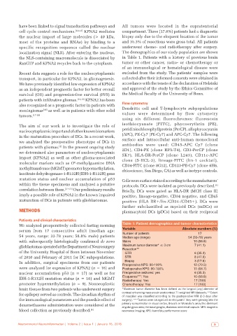
We analyzed preoperatively collected fasting morning Table 1: Patient demographics and tumor characteristics
serum from 17 consecutive adult (median age: Variable Absolute numbers (%)
17
54 years, range: 33-78 years; 58.8% male) patients Number of patients 54 (33‑78) years
Median age (range)
with subsequently histologically confirmed de novo Males 10 (58.8)
glioblastomas operated at the Department of Neurosurgery Maximum tumor diameter* ≤ 3 cm 7 (41.1)
of the University Hospital of Bonn between November Resection** 6 (35.3)
GTR
of 2010 and February of 2011 for DC subpopulations. STR 8 (47.0)
In addition, surgical specimens from our patients Biopsy 3 (17.6)
were analyzed for expression of KPNA2 (n = 16) and Preoperative KPS: 90‑100% 12 (70.5)
Postoperative KPS: 90‑100%
11 (64.7)
nuclear accumulation p53 (n = 17) as well as for Preoperative seizures: yes 6 (35.3)
IDH-1-R132H mutation status (n = 16) and MGMT Eloquence***: Yes 10 (58.8)
Radiotherapy: Yes
17 (100)
promoter hypermethylation (n = 9). Nonneoplastic Chemotherapy: Yes 17 (100)
brain tissues from two patients who underwent surgery *Maximum tumor diameter has been defined as the longest (any) diameter of
for epilepsy served as controls. The circadian rhythm of contrast enhancing mass area in postcontrast T1‑weighted MRI datasets; **Extent
of resection was classified according to the postoperative MRI (2‑3 days after
the immunological parameters and the possible effect of surgery); ***Tumors were categorized as eloquent if they were growing into the
dexamethasone administration were considered at the primary sensorimotor or visual cortex, Broca’s or Wernicke’s area/the dominant
angular gyrus area, the basal ganglia, thalamus or internal capsule. MRI: magnetic
blood collection as previously described. [4] resonance imaging; KPS: karnofsky performance score
Neuroimmunol Neuroinflammation | Volume 2 | Issue 1 | January 15, 2015 9