
Page 111 - Read Online
P. 111
irregular and often damaged, even denuded, a probable shear stress can result in activation of inflammatory
consequence of disturbed hemodynamic stress. [8] mediators, such as the master regulator of inflammation,
nuclear factor-kappaB (NF-κB). [19,20] Mechanical
Although shear stressors likely trigger the initial injury, stressors can denude the endothelium, triggering the
further degradation and disorganization of the vascular expression of chemoattractants, pro-inflammatory
wall leading to the aneurysmal growth is likely the cytokines, and cell adhesion molecules at the surface
result of an inflammatory cascade. [9-11] In general, of endothelial cells. [21] Absent from normal control
the vessel wall is transformed into a disorganized arteries, monocyte chemoattractant protein-1 (MCP-1)
array, with fragmentation/loss of the internal elastic and interleukin-8 (IL-8) are expressed in human
lamina, myointimal hyperplasia, and disorganization and experimental IAs [22] and vascular cell adhesion
of muscle fiber structure. [12-14] SMCs transition molecule-1 (VCAM-1) is expressed in the walls of
from a contractile phenotype to a pro-remodeling, human and rat model IAs. [23]
pro-inflammatory synthetic phenotype, and finally
to a dedifferentiated phenotype prior to aneurysm Macrophages and other inflammatory infiltrates
rupture. [15] Though the initial vascular injury was Numerous studies have demonstrated the presence of
from high shear stress, the cavity of the aneurysm is inflammatory cell infiltrates, particularly macrophages,
subjected to low, atheroprone-like shear stress, the in IAs. [24] In one study, inflammatory infiltrates were
type conducive to inflammatory cell adhesion and present in half of all unruptured aneurysms (10/20)
infiltration. [16] In large aneurysms (e.g. those prone versus 100% of all ruptured aneurysms (40/40). [25]
to rupture), there are often advanced atherosclerotic And in a study by Frösen et al. [26] whereby 42 ruptured
changes, phenotypically modified SMCs, lipid-laden IAs were histologically compared with 24 unruptured
macrophages, and lymphocytes. [17] IAs, infiltration of the aneurysm wall by macrophages
correlated strongly with aneurysm rupture. Macrophages
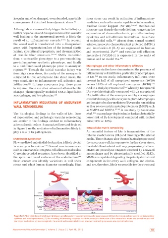
INFLAMMATORY MEDIATORS OF ANEURYSM are thought to be a key mediator of IA vascular remodeling
WALL REMODELING as they release matrix metalloproteinases (MMP) such
as MMP-9 and MMP-2. [27,28] In one study by Kanematsu
The histological findings in the walls of IAs, those et al., [29] macrophage-depleted mice had a substantially
of degeneration and pathologic vascular remodeling, lower risk of IA development compared with control
are similar to the findings evident in inflammatory mice (10% vs. 60%).
atherosclerotic lesions. Summarized here and depicted
in Figure 1 are the mediators of inflammation likely to Extracellular matrix remodeling
play a role in IA pathogenesis. An essential feature of IAs is fragmentation of the
internal elastic lamina (IEL) and thinning of the arterial
Endothelial dysfunction media. These changes alter the mechanical properties of
Flow-mediated endothelial dysfunction is likely pivotal the aneurysm wall; in response to further shear stress,
in aneurysm formation. [18] Several mechanosensors, the destabilized arterial wall may progressively balloon.
such as ion channels, integrins, cell adhesion molecules, MMPs are proteolytic enzymes secreted by activated
G-protein-coupled receptors, have been identified at macrophages and by phenotypically modified SMCs.
the apical and basal surfaces of the endothelium; [14] MMPs are capable of degrading the principal structural
these sensors can identify variations in wall shear components in the artery wall, collagen, and elastin,
stress and adapt lumen diameter accordingly. High and are, therefore, likely responsible for the structural
Figure 1: (1) Flow‑related endothelial injury; (2) triggers an inflammatory response whereby cells (macrophages) infiltrate the arterial wall and secrete pro‑inflammatory
cytokines and metalloproteinases; (3) the mounting inflammatory response results in proteolytic destruction of the extracellular matrix and smooth muscle cell
phenotypic modulation; (4) macroscopically, the arterial wall is remodeled into an aneurysm wall with progressive aneurysmal ballooning
Neuroimmunol Neuroinflammation | Volume 2 | Issue 2 | April 15, 2015 103