
Page 103 - Read Online
P. 103
the highly effective and quickly-amplified nature In PNH, for example, decreased expression of the
of the activated complement cascade, the acute complement regulators CD55 and CD59 allows for
pathogenesis of aneurysm rupture is of particular complement-mediated lysis of red blood cells in
interest. Understanding the role of complement in this a predominantly intravascular hemolysis. [10,68]
mechanism as well as the chronic processes responsible Anticomplement therapy already exists to treat many of
for aneurysm development is invaluable for future these conditions that directly result from complement
clinical endeavors. dysregulation. These include a complement component-1
(C1)-inhibitor concentrate (which inactivates C1r and
FUNCTION OF THE COMPLEMENT SYSTEM C1s and mannose-binding lectin (MBL)-associated
protein 2 (MASP 2) and is approved to treat hereditary
The complement system, a network of approximately angioedema [69,70] ), as well as eculizumab (a monoclonal
30 plasma and membrane-associated proteins, is a antibody approved for PNH and atypical hemolytic
major mediator of innate immunity, functioning in uremic syndrome [71] ). Anticomplement therapy may
cell lysis (e.g. lysis of microbes, virus-infected cells, also attenuate the damage from ischemia-reperfusion
tumor cells), inflammation, cell signaling, chemotaxis, injury. [72-74]
opsonization, and vascular effects. [54-57] In addition,
complement facilitates the adaptive immune response CLASSICAL, LECTIN, AND ALTERNATIVE
by functioning in antigen presentation, immunologic PATHWAYS
memory, and costimulation of B-cells via antigen
receptors. The presentation of “nonself” or damaged There are three recognized pathways of complement
cells leads to a cascade of events that result in the system activation: the classical, lectin, and alternative
destruction of the microbes or targeted cells and pathways [Figure 1]. The common point of each pathway
subsequent inflammation. The cascade is catalyzed by is the formation of a C3 convertase, which activates
complement components (many of which are proteases) C3 by cleaving it into C3b and C3a. [75] C3 activation
that circulate in inactive forms (zymogens) until they serves as a nidus for amplification of the complement
are activated by several mechanisms. [10] Excessive response. All three pathways eventually form C5
complement activation, however, damages healthy convertases that cleave C5 into C5a and C5b, after
tissue, and is implicated in a variety of central nervous which the C5b fragment initiates assembly of C6, C7,
system conditions (SAH, intracerebral hemorrhage, C8, and C9 into the membrane attack complex (MAC;
ischemic stroke, ischemia-reperfusion injury, and also known as the terminal complement cascade, or
multiple sclerosis [58-60] ) as well as myocardial infarctions C5b-9) which lyses the cell by forming a pore in the
and asthma. [61-64] In SAH in particular, complement lipid bilayer. [57]
activation has been associated with poorer functional
outcomes and even vasospasm. [60,65-67] The classical pathway is primarily activated by
antigen-antibody complexes. After binding to an
Dysregulation of any of the above processes, antigen, the Fc region of the antibody (typically IgM
deficiencies in the complement proteins, and activation or IgG) undergoes a conformational change that allows
by various molecules can lead to a pathological over-or it to bind to the C1q subunit of C1, a multimer that
under-activation of the complement system. These also contains C1r and C1s subunits. The C1s subunit
complement disorders [Table 1] include paroxysmal then cleaves C4 and C2, and then two of the products,
nocturnal hemoglobulinuria (PNH), hereditary C4b and C2a, associate to form the C3-convertase,
angioedema, and atypical hemolytic uremic syndrome. C4bC2a. C4bC2a also serves as the C3 convertase in
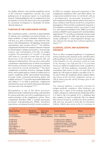
Table 1: Complement pathway disorders
Classic pathway Membrane Alternative Control proteins Others
attack complex pathway
C1q deficiency C5‑9 deficiency Factor B deficiency Factor I deficiency Serosal protease deficiency
C1r/C1s deficiency Factor D deficiency Factor H deficiency Mannose binding lectin
deficiency
C4 deficiency C4 binding protein deficiency
C2 deficiency C1 inhibitor protein deficiency
C3 deficiency Complement receptor 1‑3 deficiency
Scleroderma Paroxysmal noctural hemoglobinuria
Immunoglobulin A Leukocyte adhesion deficiency
nephropathy syndrome
Henoch-Schonlein purpura Hereditary angioedema
Membranous nephropathy Age-related macular degeneration
Systemic lupus erythematosus
Neuroimmunol Neuroinflammation | Volume 2 | Issue 2 | April 15, 2015 95