
Page 143 - Read Online
P. 143
documentation of immunohistochemical staining was expression score: 4; range: 1-12) was strongly varied
performed using an Olympus BX50 light microscope. in tumors with similar endothelial cell scores (median
expression: 3; range: 0-12). No significant correlation
Statistical analysis between tumor and endothelial cell expression scores
The semi‑quantitative TNFRSF9 scores were assigned was found [Figure 2a]. These findings point to distinct
as ordinal scaled response variables and analyzed regulatory mechanism of TNFRSF9 in melanoma and
together with nominal, ordinal, or continuous endothelial cells. Of note, in cases with an endothelial
variables. Nominal and ordinal data was analyzed cell score of > 8, no melanomas with a tumor cell score
using a contingency table followed by likelihood ratio < 4 were found.
and Pearson tests. Survival analyses were performed
using Kaplan‑Meier and multivariate analyses. In order Survival of melanoma brain metastasis patients is not
to compare survival curves, Wilcoxon and log‑rank associated with tumor necrosis factor receptor superfamily
tests were used for censored data. TNFRSF9 expression member 9 expression
levels were dichotomized at the median and referred To address the question of a potential clinicopathological
to as low or high. A significance level of alpha = 0.05
was selected for all tests. Statistical analysis was
performed using JMP 11.0.0 software (SAS Institute,
Cary, NC, USA).
RESULTS
Tumor necrosis factor receptor superfamily member 9 is
expressed on tumor and endothelial cells in melanoma a b
brain metastasis
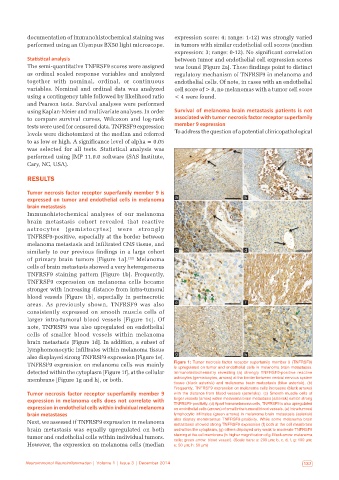
Immunohistochemical analyses of our melanoma
brain metastasis cohort revealed that reactive
astrocytes (gemistocytes) were strongly
TNFRSF9‑positive, especially at the border between
melanoma metastasis and infiltrated CNS tissue, and
similarly to our previous findings in a large cohort c d
of primary brain tumors [Figure 1a]. [12] Melanoma
cells of brain metastasis showed a very heterogeneous
TNFRSF9 staining pattern [Figure 1b]. Frequently,
TNFRSF9 expression on melanoma cells became
stronger with increasing distance from intra‑tumoral
blood vessels [Figure 1b], especially in perinecrotic
areas. As previously shown, TNFRSF9 was also e f f
consistently expressed on smooth muscle cells of
larger intra‑tumoral blood vessels [Figure 1c]. Of
note, TNFRSF9 was also upregulated on endothelial
cells of smaller blood vessels within melanoma
brain metastasis [Figure 1d]. In addition, a subset of
lymphomonocytic infiltrates within melanoma tissue
also displayed strong TNFRSF9 expression [Figure 1e]. g g h h
TNFRSF9 expression on melanoma cells was mainly Figure 1: Tumor necrosis factor receptor superfamily member 9 (TNFRSF9)
is upregulated on tumor and endothelial cells in melanoma brain metastases.
detected within the cytoplasm [Figure 1f], at the cellular Immunohistochemistry revealing (a) strongly TNFRSF9‑positive reactive
membrane [Figure 1g and h], or both. astrocytes (gemistocytes; arrows) at the border between central nervous system
tissue (black asterisk) and melanoma brain metastasis (blue asterisk). (b)
Frequently, TNFRSF9 expression on melanoma cells increases (black arrows)
Tumor necrosis factor receptor superfamily member 9 with the distance from blood vessels (asterisks). (c) Smooth muscle cells of
expression in melanoma cells does not correlate with larger vessels (arrow) within melanoma brain metastasis (asterisk) exhibit strong
TNFRSF9‑positivity. (d) Apart from melanoma cells, TNFRSF9 is also upregulated
expression in endothelial cells within individual melanoma on endothelial cells (arrows) of small intra‑tumoral blood vessels. (e) Intra‑tumoral
brain metastases lymphocytic infiltrates (green arrows) in melanoma brain metastasis (asterisk)
Next, we assessed if TNFRSF9 expression in melanoma also display membranous TNFRSF9‑positivity. While some melanoma brain
metastases showed strong TNFRSF9 expression (f) both at the cell membrane
brain metastasis was equally upregulated on both and within the cytoplasm, (g) others displayed only weak to moderate TNFRSF9
tumor and endothelial cells within individual tumors. staining at the cell membrane (h: higher magnification of g. Black arrow: melanoma
cells; green arrow: blood vessel). (Scale bars: a: 200 µm; b, c, d, f, g: 100 µm;
However, the expression on melanoma cells (median e: 50 µm; h: 50 µm)
Neuroimmunol Neuroinflammation | Volume 1 | Issue 3 | December 2014 137