
Page 142 - Read Online
P. 142
Furthermore, tumor‑infiltrating lymphocytes (TIL) cases with > 10 metastases in one patient, the number
which have been stimulated with agonistic TNFRSF9 of metastases was set to 10 for statistical analysis.
antibodies show a stronger antitumor effect as well as Patient age at surgery and overall survival after surgery
prolonged survival. Therefore, stimulating TNFRSF9 were registered.
[4]
by the use of agonistic antibodies has been proposed
as an additional immunotherapeutic approach in Immunohistochemistry
cancer treatment‑especially for melanoma, which For immunohistochemistry, a mouse monoclonal
represents one of the most immunogenic tumors‑and antihuman TNFRSF9 antibody (dilution 1:40; clone S16,
has already entered clinical trials. [5,6] An alternative Novocastra/Leica Microsystems, Germany) was used as
[12]
approach uses genetically modified human T‑cells previously published. Tissue labeling was performed
[7]
to express higher TNFRSF9 levels. TNFRSF9 using the Discovery XT immunohistochemistry
stimulation has been suggested as a treatment for system (Ventana Medical Systems, France). A cell
metastatic cancers. However, to date there is conditioning pretreatment was performed for 68 min
[8]
only poor data about the distribution of TNFRSF9 followed by a 4 min blocking step with inhibitor
in brain metastases, which still constitute one of D. The primary antibody was applied for 32 min,
the most deleterious clinical conditions in tumor followed by secondary antibody (Discovery Universal
patients. [9,10] This data is of importance since Secondary Antibody) for 32 min. After washing
preliminary clinical studies targeting TNFRSF9 steps, a blocking step with blocker D for 4 min and a
have already been stopped due to considerable side 16 min incubation with one drop of SA‑HRP D were
effects. [11] In a previous study, our group showed performed. For diaminobenzidine (DAB) visualization,
that TNFRSF9 was strongly upregulated by reactive the sections were incubated with one drop of DAB D
astrocytes (so‑called gemistocytes) in primary central and one drop of DAB H O D for 8 min, followed by
2
2
nervous system (CNS) tumors, whereas both brain a copper enhancer (Copper D, all Ventana Medical
tumor cells and TIL were mainly TNFRSF9‑negative. [12] Systems, Tucson, AZ, USA) for 4 min. Specimens
Since most studies have only focused on TNFRSF9 were washed, counterstained with hematoxylin and
expression on hematopoietic cells, there is an urgent bluing reagent, and mounted. In addition, our cohort
need to decipher TNFRSF9 expression on other cell was immunohistochemically assessed for BRAF V600E
types and different microenvironmental conditions mutations using mouse monoclonal IgG2a antihuman
in vivo in more detail. Of note, a recent animal study BRAF V600E (dilution 1:100; clone VE1, Spring
discovered that TNFRSF9 is also expressed in neural Bioscience). Images were analyzed and recorded on
stem cells, in which it induced cell death. [13] The an Olympus BX‑50 microscope (Olympus, Germany).
expression of TNFRSF9 on cell types other than
hematopoietic cells might be at least partly responsible Scoring
for side effects in preliminary clinical trials targeting Tumor necrosis factor receptor superfamily member
TNFRSF9. Therefore, the aim of our current study was 9 expression was separately assessed in both tumor
to define the cellular source of TNFRSF9 expression and endothelial cells by taking staining intensity and
in melanoma brain metastasis, in order to assess frequency into account, using a previously established
the suitability of an anti‑TNFRSF9 treatment in this protocol. [14,15] The semi‑quantitative scores consist of
detrimental clinical condition. a frequency score ranging from 0 to 4 (0 = 0‑1%,
1 = 2-10%, 2 = 11-25%, 3 = 26-50%, and 4 ≥ 50% of
METHODS all cells showing positive nuclear staining). Likewise,
intensity was recorded in a similar semi‑quantitative
Patient data approach as follows: 0 = no staining, 1 = weak staining,
The use of human tissue from cases of melanoma 2 = moderate staining, and 3 = strong staining. The
brain metastasis and the respective clinical data was scores for staining intensity and frequency were
approved by the ethical committee of the Eberhard multiplied together, so that the final expression cell
Karls University of Tübingen and Tübingen University score reflected both. The evaluation and photographic
Hospital (project no. 408/2013BO2). Our cohort
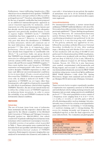
consisted of 78 patients suffering from melanoma brain Table 1: Patient data
metastases which underwent neurosurgical resection. Characteristic Data
A detailed overview of our patient cohort is provided Patient age, median (range) 60 (20‑83)
in Table 1. Tissue microarrays were constructed from Sex, male/female 47/31
formalin‑fixed and paraffin‑embedded tumor samples Number of brain metastases, median (range) 1 (1‑10)
for immunohistochemical analysis of TNFRSF9 Tumor size in mm, median (range) 28 (5‑61)
38/40
BRAF V600E mutation assessed by IHC, yes/no
expression. Brain magnetic resonance imaging data was Survival in days, median (range) 177 (17‑4166)
analyzed for metastasis size (diameter) and number. In IHC: Immunohistochemistry, BRAF: proto-oncogene B-Raf
136 Neuroimmunol Neuroinflammation | Volume 1 | Issue 3 | December 2014