
Page 144 - Read Online
P. 144
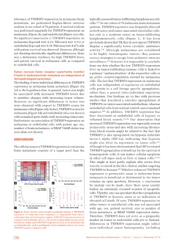
relevance of TNFRSF9 expression in melanoma brain   typically assessed tumor‑infiltrating lymphomonocytic
           metastasis, we performed Kaplan‑Meier survival     cells. [2,3]  In our cohort of 78 melanoma brain metastasis
           analysis in our cohort of 78 patients. A survival analysis   patients, TNFRSF9 expression was frequently detected
           was performed separately for TNFRSF9 expression on   on both tumor and tumor‑associated endothelial cells,
           melanoma [Figure 2b] and endothelial [Figure 2c] cells.   but only to a moderate extent on tumor‑infiltrating
           No significant association of TNFRSF9 expression on   lymphomonocytic cells  [Figure  1]. It has been
           melanoma (log‑rank test: 0.23; Wilcoxon test: 0.31) or   previously shown that TIL that do not express TNFRSF9
           endothelial (log‑rank test: 0.39; Wilcoxon test: 0.67) cells   display a significantly lower cytolytic antitumor
           with patient survival was observed. However, although   activity. [16]  Although melanomas are considered
           not showing statistically significant differences, there   to be highly immunogenic tumors, they possess
           was a dichotomic tendency for high TNFRSF9 levels   various strategies to escape from antitumor immune
           and patient survival in melanoma cells as compared   surveillance. [17]  However, it is impossible to conclude
           to endothelial cells.                              from our data whether the low TNFRSF9 expression
                                                              level on tumor‑infiltrating immune cells is linked to
           Tumor necrosis factor receptor superfamily member   a primary “underactivation” of the respective cells or
           9 levels in melanoma brain metastasis are independent of   an active counter‑regulation exerted by melanoma
           clinicopathological parameters
           The finding of intra‑individual differences in TNFRSF9   cells. The fact that TNFRSF9 expression on melanoma
           expression in melanoma brain metastasis [Figure 1b]   cells was independent of expression on endothelial
           led to the hypothesis that, in general, tumor size might   cells points to a cell lineage specific upregulation,
           be associated with increased TNFRSF9 levels due    rather than a general intra‑individual regulatory
           to nutritive changes with increasing tumor volume.   mechanism. Our findings are in line with previous
           However, no significant differences in tumor size   studies that described a selective upregulation of
           were observed with respect to TNFRSF9 scores for   TNFRSF9 on tumor‑associated endothelium, whereas
           melanoma cells [Figure 2d]. In fact, TNFRSF9 scores for   endothelial cells from normal control cases remained
           melanoma [Figure 2d] and endothelial (data not shown)   negative. [18]  In addition, TNFRSF9 expression has
           cells remained quite stable with increasing tumor size.   been discovered on endothelial cells of hypoxic or
           Furthermore, no association of TNFRSF9 expression on   inflamed blood vessels. [19,20]  Our observation that
           melanoma or endothelial cells with patient age, sex,   increased TNFRSF9 expression was especially seen in
           number of brain metastases, or BRAF V600E status was   perinecrotic areas‑and also with increasing distance
           seen (data not shown).                             from blood vessels‑might be related to the fact that
                                                              TNFRSF9 is also upregulated via hypoxia inducible
           DISCUSSION                                         factor‑1 alpha  (HIF‑1α), indicating that hypoxia
                                                              might also drive its expression on tumor cells. [21]
           The cellular source of TNFRSF9 expression in melanoma   Although it has been demonstrated that HIF1α‑related
           brain metastasis consists of a larger pool than the   TNFRSF9 upregulation is beneficial for the survival of
                                                              hematopoietic cells, it can induce cellular apoptosis
                                                              in other cell types such as liver or tumor cells. [22‑24]
                                                              This might at least partly explain why severe liver
                                                              toxicity occurred in the first clinical studies targeting
                                                              TNFRSF9 in humans.  [25]  Whether enhanced TNFRSF9
                                                              expression in perinecrotic areas in melanoma brain
                                                              metastasis is beneficial or detrimental to the tumor
                                                              remains an open question. However, a conclusion
           a                         b
                                                              by analogy can be made, since these areas usually
                                                              harbor an extremely elevated number of apoptotic
                                                              cells. Thereby, one can speculate that the upregulation
                                                              of TNFRSF9 in hypoxic areas is an indication of
                                                              elevated cell death. Of note, TNFRSF9 expression on
                                                              either tumor or endothelial cells was not associated
           c                       d                          with age, sex, patient survival, size or number of
           Figure 2: Tumor necrosis factor receptor superfamily member 9 (TNFRSF9)   brain metastases, or BRAF V600E expression status.
           expression in melanoma brain metastasis is independent from clinico‑  Therefore, TNFRSF9 does not serve as a prognostic
           pathological data. (a) Contingency analysis of TNFRSF9 expression scores for
           melanoma and endothelial cells (n = 78). (b and c) Kaplan‑Meier survival curves   marker on tumor or endothelial cells per se. Instead,
           stratified by median split of TNFRSF9 expression scores for (b) melanoma and   differences in TNFRSF9 expression might reflect
           (c) endothelial cells. (d) Box‑plot diagram of brain metastasis size (in mm) versus
           TNFRSF9 score for melanoma cells                   inter‑individual tumor heterogeneity, including


            138                                             Neuroimmunol Neuroinflammation | Volume 1 | Issue 3 | December 2014