
Page 307 - Read Online
P. 307
Page 6 of 12 Torabinia et al. Mini-invasive Surg 2021;5:32 https://dx.doi.org/10.20517/2574-1225.2021.63
Table 1. Dice, Precision, and Recall metrics evaluation of catheter tip's radiopaque marker testing set for segmentation task by U-
Net model
Method Catheter tip's radiopaque marker segmentation
Indexes Dice coefficient Binary cross-entropy Intersection over Union
Deep learning 0.8457 0.3512 0.8367
U-Net model
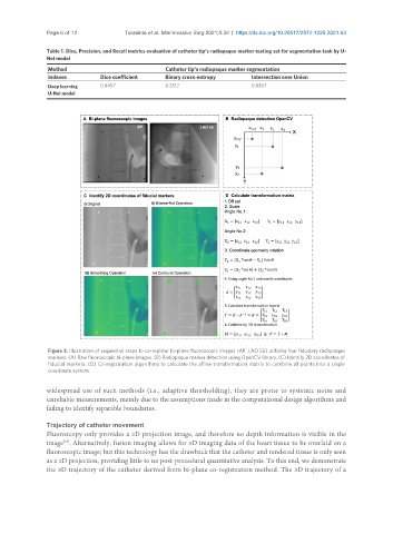
Figure 3. Illustration of sequential steps to co-register bi-plane fluoroscopic images (AP, LAO 55) utilizing four fiduciary radiopaque
markers. (A) Raw fluoroscopic bi-plane images. (B) Radiopaque marker detection using OpenCV library. (C) Identify 2D coordinates of
fiducial markers. (D) Co-registration algorithms to calculate the affine transformation matrix to combine all points into a single
coordinate system.
widespread use of such methods (i.e., adaptive thresholding), they are prone to systemic noise and
unreliable measurements, mainly due to the assumptions made in the computational design algorithms and
failing to identify separable boundaries.
Trajectory of catheter movement
Fluoroscopy only provides a 2D projection image, and therefore no depth information is visible in the
image . Alternatively, fusion imaging allows for 3D imaging data of the heart tissue to be overlaid on a
[57]
fluoroscopic image; but this technology has the drawback that the catheter and rendered tissue is only seen
as a 2D projection, providing little to no post-procedural quantitative analysis. To this end, we demonstrate
the 3D trajectory of the catheter derived from bi-plane co-registration method. The 3D trajectory of a