
Page 304 - Read Online
P. 304
Torabinia et al. Mini-invasive Surg 2021;5:32 https://dx.doi.org/10.20517/2574-1225.2021.63 Page 3 of 12
Methodology
A schematic of the proposed training system is shown in Figure 1, where a physician conducts a mock
catheterization procedure using a bi-plane C-arm X-ray fluoroscopy machine on a patient-specific 3D
printed model. The proposed image tracking aims to detect and co-register the catheter's 3D position and
provide a 3D trajectory as quantitative feedback. Different features that are utilized for our proposed
tracking system are described in detail in the following subsections, which are in the order by which this
process is conducted.
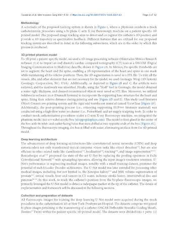
3D printed phantom model
To 3D print a patient-specific model, we used a 3D image processing software (Materialize Mimics Research
software 21.0) to import an end-diastolic cardiac computed tomography (CT) scan as a DICOM (Digital
Imaging Communication in Medicine) data file, shown in Figure 2A. In Mimics, the specific thresholds are
set to segment the heart and the spine, enabling a 3D representation of the heart and spine in one mask
while maintaining all the relative positions. Then, the 3D segmentation is saved as a STL file. To trim all the
vessels, ribs, and other elements that are not necessary for the model, we used Geomagic Wrap (3D Systems
Geomagic Corporation, NC, USA). Additionally, as depicted in Figure 2B and C, the artifacts were
removed, and the meshwork was smoothed. Finally, using the “Shell” tool in Geomagic, the model obtained
a water-tight thickness, and cleaned reconstructed objects were saved as STL files. Moreover, we utilized
Solidworks software 2018 (Dassault Systems) to incorporate the supporting base structure for the heart and
spine, fixing their relative distance during printing and use [Figure 2D and E]. This study used Stratasys
Object Connex 260 printing system and the rigid and translucent material named VeroClear [Figure 2F].
Additionally, the post-printing process (i.e., removing supporting SUP705 Stratasys material) was
conducted using a high-flow water jet cleaner (i.e., Powerblast) and art supply sculpting tools. In order to
conduct mock catheterization procedures under a C-arm X-ray fluoroscopy machine, we integrated the
phantom model into a 5-sided acrylic box (shoppopdisplays.com). The model is then glued in the center of
the box with its inlet- and outlet-facing holes that were drilled at two opposite ends of the box [Figure 2G].
Throughout the fluoroscopic imaging, the box is filled with water, eliminating artifacts from the 3D printed
model.
Deep learning architecture
The advancement of deep learning architectures like convolutional neural networks (CNN) and deep
[36]
autoencoders not only transformed typical computer vision tasks like object detection , but are also
efficient in other related tasks like classification , localization , tracking , and image segmentation [40,41] .
[37]
[38]
[39]
[41]
Ronneberger et al. proposed the state-of-the-art U-Net by replacing the pooling operators in Fully
Convolutional Network with upsampling operators, allowing the input image's resolution retention. U-
[42]
Net's performance in segmenting medical images, notably with a small training dataset, promises the
potential of such Encoder-Decoder architecture. The U-Net model was later extended for processing other
medical images, including, but not limited to, the Xenopus kidney and MRI volume segmentation of
[43]
[44]
prostate , retinal vessels, liver and tumors in CT scans, ischemic stroke lesion, intervertebral disc and
pancreas [45-52] . In this work, to track the catheter's position from the bi-plane fluoroscopic images, we
primarily leveraged the U-Net model to detect a radiopaque marker at the tip of the catheter. The details of
implementation and framework will be discussed in the following sections.
Collection and preparation of datasets
All fluoroscopic images for training the deep learning U-Net model were acquired during the mock
procedures in the catheterization lab at New York-Presbyterian Hospital. The datasets comprise 300 paired
bi-plane images pertaining to the maneuvering of a catheter (OSCAR Deflectable Steerable Guiding Sheath,
Destino™ Twist) within the patient-specific 3D printed model. The datasets were divided into 3 parts: (1)