
Page 305 - Read Online
P. 305
Page 4 of 12 Torabinia et al. Mini-invasive Surg 2021;5:32 https://dx.doi.org/10.20517/2574-1225.2021.63
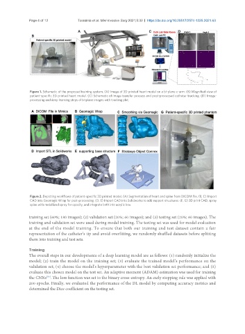
Figure 1. Schematic of the proposed training system. (A) Image of 3D printed heart model on a bi-plane c-arm. (B) Magnified view of
patient-specific 3D printed heart model. (C) Schematic of image transfer process and post-processed catheter tracking. (D) Image-
processing and deep learning steps of bi-plane images with tracking plot.
Figure 2. Depicting workflows of patient-specific 3D printed model. (A) Segmentation of heart and spine from DICOM file. (B, C) Import
CAD into Geomagic Wrap for post-processing. (D, E) Import CAD into Solidworks to add support structures. (F, G) 3D print CAD, spray
spine with metallized spray for opacity, and integrate both into acrylic box.
training set (60%; 180 images); (2) validation set (20%; 60 images); and (3) testing set (20%; 60 images). The
training and validation set were used during model training. The testing set was used for model evaluation
at the end of the model training. To ensure that both our training and test dataset contain a fair
representation of the catheter’s tip and avoid overfitting, we randomly shuffled datasets before splitting
them into training and test sets.
Training
The overall steps in our developments of a deep learning model are as follows: (1) randomly initialize the
model; (2) train the model on the training set; (3) evaluate the trained model’s performance on the
validation set; (4) choose the model’s hyperparameter with the best validation set performance; and (5)
evaluate this chosen model on the test set. An adaptive moment (ADAM) estimation was used for training
[53]
the CNNs . The loss function was set to the binary cross-entropy. An early stopping rule was applied with
200 epochs. Finally, we evaluated the performance of the DL model by computing accuracy metrics and
determined the Dice coefficient on the testing set.