
Page 310 - Read Online
P. 310
Torabinia et al. Mini-invasive Surg 2021;5:32 https://dx.doi.org/10.20517/2574-1225.2021.63 Page 9 of 12
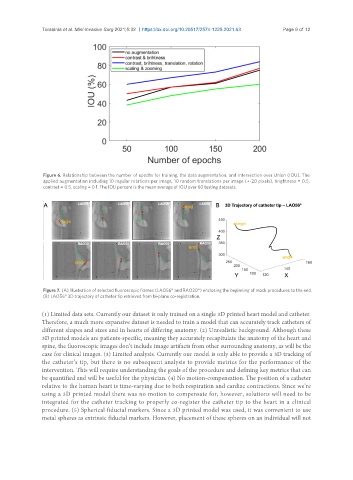
Figure 6. Relationship between the number of epochs for training, the data augmentation, and Intersection over Union (IOU). The
applied augmentation including 10 regular rotations per image, 10 random translations per image (+-20 pixels), brightness = 0.5,
contrast = 0.5, scaling = 0.1. The IOU percent is the mean average of IOU over 60 testing datasets.
Figure 7. (A) Illustration of selected fluoroscopic frames (LAO56° and RAO30°) enclosing the beginning of mock procedures to the end.
(B) LAO56° 3D trajectory of catheter tip retrieved from bi-plane co-registration.
(1) Limited data sets. Currently our dataset is only trained on a single 3D printed heart model and catheter.
Therefore, a much more expansive dataset is needed to train a model that can accurately track catheters of
different shapes and sizes and in hearts of differing anatomy. (2) Unrealistic background. Although these
3D printed models are patients-specific, meaning they accurately recapitulate the anatomy of the heart and
spine, the fluoroscopic images don’t include image artifacts from other surrounding anatomy, as will be the
case for clinical images. (3) Limited analysis. Currently our model is only able to provide a 3D tracking of
the catheter’s tip, but there is no subsequent analysis to provide metrics for the performance of the
intervention. This will require understanding the goals of the procedure and defining key metrics that can
be quantified and will be useful for the physician. (4) No motion-compensation. The position of a catheter
relative to the human heart is time-varying due to both respiration and cardiac contractions. Since we’re
using a 3D printed model there was no motion to compensate for, however, solutions will need to be
integrated for the catheter tracking to properly co-register the catheter tip to the heart in a clinical
procedure. (5) Spherical fiducial markers. Since a 3D printed model was used, it was convenient to use
metal spheres as extrinsic fiducial markers. However, placement of these spheres on an individual will not