
Page 170 - Read Online
P. 170
Avery et al. Mini-invasive Surg 2021;5:17 https://dx.doi.org/10.20517/2574-1225.2021.05 Page 7 of 18
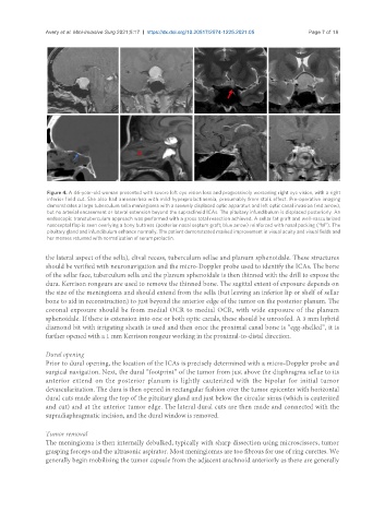
Figure 4. A 46-year-old woman presented with severe left eye vision loss and progressively worsening right eye vision, with a right
inferior field cut. She also had amenorrhea with mild hyperprolactinemia, presumably from stalk effect. Pre-operative imaging
demonstrates a large tuberculum sella meningioma with a severely displaced optic apparatus and left optic canal invasion (red arrow),
but no arterial encasement or lateral extension beyond the supraclinoid ICAs. The pituitary infundibulum is displaced posteriorly. An
endoscopic transtuberculum approach was performed with a gross total resection achieved. A sellar fat graft and well-vascularized
nasoseptal flap is seen overlying a bony buttress (posterior nasal septum graft; blue arrow) reinforced with nasal packing (“M”). The
pituitary gland and infundibulum enhance normally. The patient demonstrated marked improvement in visual acuity and visual fields and
her menses returned with normalization of serum prolactin.
the lateral aspect of the sella), clival recess, tuberculum sellae and planum sphenoidale. These structures
should be verified with neuronavigation and the micro-Doppler probe used to identify the ICAs. The bone
of the sellar face, tuberculum sella and the planum sphenoidale is then thinned with the drill to expose the
dura. Kerrison rongeurs are used to remove the thinned bone. The sagittal extent of exposure depends on
the size of the meningioma and should extend from the sella (but leaving an inferior lip or shelf of sellar
bone to aid in reconstruction) to just beyond the anterior edge of the tumor on the posterior planum. The
coronal exposure should be from medial OCR to medial OCR, with wide exposure of the planum
sphenoidale. If there is extension into one or both optic canals, these should be unroofed. A 3 mm hybrid
diamond bit with irrigating sheath is used and then once the proximal canal bone is “egg-shelled”, it is
further opened with a 1 mm Kerrison rongeur working in the proximal-to-distal direction.
Dural opening
Prior to dural opening, the location of the ICAs is precisely determined with a micro-Doppler probe and
surgical navigation. Next, the dural “footprint” of the tumor from just above the diaphragma sellae to its
anterior extend on the posterior planum is lightly cauterized with the bipolar for initial tumor
devascularization. The dura is then opened in rectangular fashion over the tumor epicenter with horizontal
dural cuts made along the top of the pituitary gland and just below the circular sinus (which is cauterized
and cut) and at the anterior tumor edge. The lateral dural cuts are then made and connected with the
supradiaphragmatic incision, and the dural window is removed.
Tumor removal
The meningioma is then internally debulked, typically with sharp dissection using microscissors, tumor
grasping forceps and the ultrasonic aspirator. Most meningiomas are too fibrous for use of ring curettes. We
generally begin mobilizing the tumor capsule from the adjacent arachnoid anteriorly as there are generally