
Page 166 - Read Online
P. 166
Avery et al. Mini-invasive Surg 2021;5:17 https://dx.doi.org/10.20517/2574-1225.2021.05 Page 3 of 18
assisted endonasal approach. Since 2009, we have transitioned to a fully endoscopic endonasal approach
while gaining more experience with the supraorbital route for parasellar tumors and have reversed the ratio
to 61% endoscopic endonasal and 39% supraorbital route [2,10,28-30] .
There are several major advantages of the endonasal route. The natural nasal corridor provides a direct
trajectory to the tuberculum sellae and posterior planum, facilitating tumor removal with minimal brain
manipulation. The meningioma lies between the surgeon and critical structures such as the optic nerves,
optic chiasm and ICAs, thereby minimizing risk of iatrogenic injury. Medial optic canal decompression can
be safely and effectively performed when optic canal invasion is present. Devascularization is accomplished
early in surgery by interrupting the dural blood supply during the approach. Finally, adjacent hyperostotic
bone is readily removed en route to the meningioma.
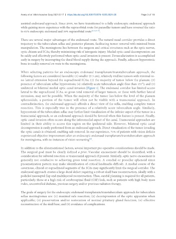
When selecting patients for an endoscopic endonasal transplanum/transtuberculum approach, the
following factors are considered favorable: (1) smaller (≤ 3 cm), relatively midline tumors with minimal-to-
no lateral extension beyond the supraclinoid ICAs; (2) the majority of tumor below the planum; (3)
presence of tuberculum sellae hyperostosis; (4) relatively acute tuberculum angle (less than 135°); and (5)
unilateral or bilateral medial optic canal invasion [Figure 1]. The endonasal corridor has limited access
lateral to the supraclinoid ICAs, so gross total removal of larger tumors, or those with further lateral
extension, may not be possible. When the majority of the tumor lies below the level of the planum
sphenoidale, a portion of the tumor will often not be visible with a transcranial approach. In
contradistinction, the endonasal approach affords a direct view of the sella, enabling complete tumor
resection. This is especially true in the presence of a relatively acute tuberculum angle. Similarly,
hyperostosis of the tuberculum sellae may further limit visualization of the inferior aspect of the tumor in a
transcranial approach, so an endonasal approach should be favored when this feature is present. Finally,
optic canal invasion often occurs along the inferomedial aspect of the canal. Transcranial approaches are
limited in their ability to access this region on the ipsilateral side. However, bilateral optic canal
decompression is easily performed from an endonasal approach. Direct visualization of the tumor invading
the optic canals is obtained, enabling safe removal. In our experience, 71% of patients with vision deficits
experienced objective improvement after an endoscopic endonasal transplanum/transtuberculum approach
[30]
for meningioma, with no instances of vision worsening .
In addition to the aforementioned factors, several important pre-operative considerations should be made.
The surgical goal must be clearly defined a prior. Vascular encasement should be identified, with a
consideration for subtotal resection or transcranial approach if present. Similarly, optic nerve encasement is
generally not conducive to achieving gross total resection. A conchal or presellar sphenoid sinus
pneumatization pattern may make identification of critical landmarks difficult. A medial course of the
cavernous, clinoid or supraclinoid segments of the ICAs may significantly limit the surgical corridor. The
endonasal approach creates a large dural defect requiring a robust skull base reconstruction, ideally with a
pedicled nasoseptal flap and multilayered reconstruction. Thus, careful planning is required for all patients,
particularly those at a high risk of cerebrospinal fluid (CSF) leak, such as patients with high body mass
index, uncontrolled diabetes, previous surgery and/or previous radiation therapy.
The goals of surgery for the endoscopic endonasal transplanum/transtuberculum approach for tuberculum
sellae meningiomas are: (1) maximal safe resection; (2) decompression of the optic apparatus when
applicable; (3) preservation and/or restoration of normal pituitary gland function; (4) effective
reconstruction of the skull base; and (5) avoidance of complications.