
Page 169 - Read Online
P. 169
Page 6 of 18 Avery et al. Mini-invasive Surg 2021;5:17 https://dx.doi.org/10.20517/2574-1225.2021.05
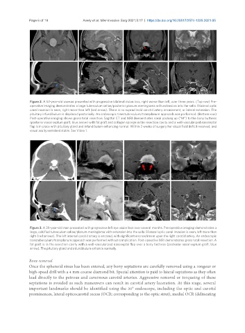
Figure 2. A 52-year-old woman presented with progressive bilateral vision loss, right worse than left, over three years. (Top row) Pre-
operative imaging demonstrates a large tuberculum sellae/posterior planum meningioma with extension into the sella. Bilateral optic
canal invasion is seen, right more than left (red arrow). There is no supraclinoid carotid artery encasement, or lateral extension. The
pituitary infundibulum is displaced posteriorly. An endoscopic transtuberculum/transplanum approach was performed. (Bottom row)
Post-operative imaging shows gross total resection. Sagittal CT and MRI demonstrates nasal packing up (“M”) to the bony buttress
(posterior nasal septum graft; blue arrow) with fat graft and collagen sponge in the resection cavity and a well-vascularized nasoseptal
flap is in place with pituitary gland and infundibulum enhancing normal. Within 2 weeks of surgery her visual field deficit resolved, and
visual acuity remained stable. See Video 1.
Figure 3. A 39-year-old man presented with progressive left eye vision loss over several months. Pre-operative imaging demonstrates a
large, calcified tuberculum sellae/planum meningioma with extension into the sella. Bilateral optic canal invasion is seen, left more than
right (red arrows). The left internal carotid artery is encased, with significant encroachment upon the right carotid artery. An endoscopic
transtuberculum/transplanum approach was performed without complication. Post-operative MRI demonstrates gross total resection. A
fat graft is in the resection cavity with a well-vascularized nasoseptal flap over a bony buttress (posterior nasal septum graft; blue
arrow). The pituitary gland and infundibulum enhance normally.
Bone removal
Once the sphenoid sinus has been entered, any bony septations are carefully removed using a rongeur or
high-speed drill with a 4 mm course diamond bit. Special attention is paid to lateral septations as they often
lead directly to the petrous and cavernous carotid arteries. Aggressive removal or torqueing of these
septations is avoided as such maneuvers can result in carotid artery laceration. At this stage, several
important landmarks should be identified using the 30° endoscope, including the optic and carotid
prominences, lateral opticocarotid recess (OCR; corresponding to the optic strut), medial OCR (delineating