
Page 715 - Read Online
P. 715
Page 8 of 12 Masiero et al. Mini-invasive Surg 2020;4:71 I http://dx.doi.org/10.20517/2574-1225.2020.56
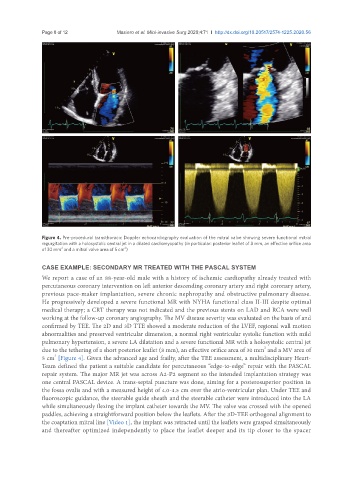
Figure 4. Pre-procedural transthoracic Doppler echocardiography evaluation of the mitral valve showing severe functional mitral
regurgitation with a holosystolic central jet in a dilated cardiomyopathy (in particular: posterior leaflet of 8 mm, an effective orifice area
2
2
of 30 mm and a mitral valve area of 5 cm )
CASE EXAMPLE: SECONDARY MR TREATED WITH THE PASCAL SYSTEM
We report a case of an 88-year-old male with a history of ischemic cardiopathy already treated with
percutaneous coronary intervention on left anterior descending coronary artery and right coronary artery,
previous pace-maker implantation, severe chronic nephropathy and obstructive pulmonary disease.
He progressively developed a severe functional MR with NYHA functional class II-III despite optimal
medical therapy; a CRT therapy was not indicated and the previous stents on LAD and RCA were well
working at the follow-up coronary angiography. The MV disease severity was evaluated on the basis of and
confirmed by TEE. The 2D and 3D TTE showed a moderate reduction of the LVEF, regional wall motion
abnormalities and preserved ventricular dimension, a normal right ventricular systolic function with mild
pulmonary hypertension, a severe LA dilatation and a severe functional MR with a holosystolic central jet
2
due to the tethering of a short posterior leaflet (8 mm), an effective orifice area of 30 mm and a MV area of
2
5 cm [Figure 4]. Given the advanced age and frailty, after the TEE assessment, a multidisciplinary Heart-
Team defined the patient a suitable candidate for percutaneous “edge-to-edge” repair with the PASCAL
repair system. The major MR jet was across A2-P2 segment so the intended implantation strategy was
one central PASCAL device. A trans-septal puncture was done, aiming for a posterosuperior position in
the fossa ovalis and with a measured height of 4.0-4.5 cm over the atrio-ventricular plan. Under TEE and
fluoroscopic guidance, the steerable guide sheath and the steerable catheter were introduced into the LA
while simultaneously flexing the implant catheter towards the MV. The valve was crossed with the opened
paddles, achieving a straightforward position below the leaflets. After the 3D-TEE orthogonal alignment to
the coaptation mitral line [Video 1], the implant was retracted until the leaflets were grasped simultaneously
and thereafter optimized independently to place the leaflet deeper and its tip closer to the spacer