Page 711 - Read Online
P. 711
Page 4 of 12                                    Masiero et al. Mini-invasive Surg 2020;4:71  I  http://dx.doi.org/10.20517/2574-1225.2020.56

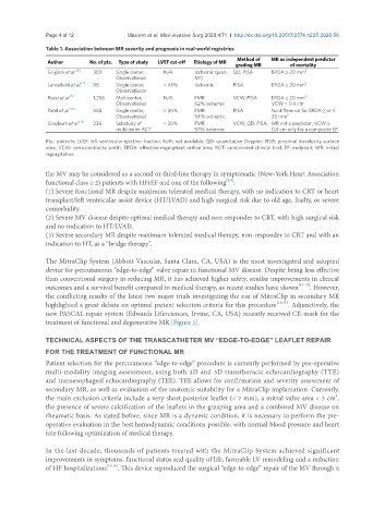
               Table 1. Association between MR severity and prognosis in real-world registries
                                                                          Method of   MR as independent predictor
                Author       No. of pts.  Type of study  LVEF cut-off  Etiology of MR
                                                                          grading MR       of mortality
                Grigioni et al. [6]  303  Single center,   N/A  Ischemic (post- QD, PISA  EROA ≥ 20 mm 2
                                     Observational           MI)
                Lancellotti et al. [7]  98  Single center,   < 45%  Ischemic  PISA   EROA ≥ 20 mm 2
                                     Observational
                Rossi et al. [5]  1,256  Multicenter,   N/A  FMR         VCW, PISA   EROA ≥ 20 mm 2
                                     Observational           62% ischemic            VCW > 0.4 cm
                Patel et al. [10]  558  Single center,   < 35%  FMR      PISA        No difference for EROA ≥ or <
                                     Observational           54% ischemic            20 mm 2
                Grayburn et al. [11]  336  Substudy of   < 35%  FMR      VCW, QD, PISA MR not a predictor; VCW ≥
                                     multicenter RCT         57% ischemic            0.4 cm only for a composite EP

               Pts.: patients; LVEF: left ventricular ejection fraction; N/A: not available; QD: quantitative Doppler; PISA: proximal isovelocity surface
               area; VCW: vena contracta width; EROA: effective regurgitant orifice area; RCT: randomized clinical trial; EP: endpoint; MR: mitral
               regurgitation

               the MV may be considered as a second or third-line therapy in symptomatic (New-York Heart Association
               functional class ≥ 2) patients with HFrEF and one of the following :
                                                                       [1,9]
               (1) Severe functional MR despite maximum tolerated medical therapy, with no indication to CRT or heart
               transplant/left ventricular assist device (HT/LVAD) and high surgical risk due to old age, frailty, or severe
               comorbidity.
               (2) Severe MV disease despite optimal medical therapy and non-responder to CRT, with high surgical risk
               and no indication to HT/LVAD.
               (3) Severe secondary MR despite maximum tolerated medical therapy, non-responder to CRT and with an
               indication to HT, as a “bridge therapy”.

               The MitraClip System (Abbott Vascular, Santa Clara, CA, USA) is the most investigated and adopted
               device for percutaneous “edge-to-edge” valve repair in functional MV disease. Despite being less effective
               than conventional surgery in reducing MR, it has achieved higher safety, similar improvements in clinical
               outcomes and a survival benefit compared to medical therapy, as recent studies have shown [13-15] . However,
               the conflicting results of the latest two major trials investigating the use of MitraClip in secondary MR
               highlighted a great debate on optimal patient selection criteria for this procedure [14,15] . Adjunctively, the
               new PASCAL repair system (Edwards Lifesciences, Irvine, CA, USA) recently received CE-mark for the
               treatment of functional and degenerative MR [Figure 2].


               TECHNICAL ASPECTS OF THE TRANSCATHETER MV “EDGE-TO-EDGE” LEAFLET REPAIR
               FOR THE TREATMENT OF FUNCTIONAL MR
               Patient selection for the percutaneous “edge-to-edge” procedure is currently performed by pre-operative
               multi-modality imaging assessment, using both 2D and 3D transthoracic echocardiography (TTE)
               and transesophageal echocardiography (TEE). TEE allows for confirmation and severity assessment of
               secondary MR, as well as evaluation of the anatomic suitability for a MitraClip implantation. Currently,
               the main exclusion criteria include a very short posterior leaflet (< 7 mm), a mitral valve area < 3 cm ,
                                                                                                         2
               the presence of severe calcification of the leaflets in the grasping area and a combined MV disease on
               rheumatic basis. As stated before, since MR is a dynamic condition, it is necessary to perform the pre-
               operative evaluation in the best hemodynamic conditions possible, with normal blood pressure and heart
               rate following optimization of medical therapy.


               In the last decade, thousands of patients treated with the MitraClip System achieved significant
               improvements in symptoms, functional status and quality of life, favorable LV remodeling and a reduction
               of HF hospitalizations [13,16] . This device reproduced the surgical “edge-to-edge” repair of the MV through a

