
Page 716 - Read Online
P. 716
Masiero et al. Mini-invasive Surg 2020;4:71 I http://dx.doi.org/10.20517/2574-1225.2020.56 Page 9 of 12
A B
C D
E F
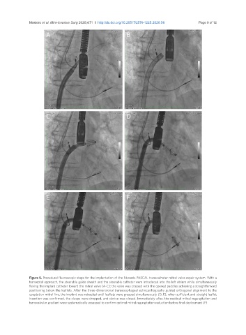
Figure 5. Procedural fluoroscopic steps for the implantation of the Edwards PASCAL transcatheter mitral valve repair system. With a
transeptal approach, the steerable guide sheath and the steerable catheter were introduced into the left atrium while simultaneously
flexing the implant catheter toward the mitral valve (A-C); the valve was crossed with the opened paddles achieving a straightforward
positioning below the leaflets. After the three-dimensional transesophageal echocardiography guided orthogonal alignment to the
coaptation mitral line, the implant was retracted until leaflets were grasped simultaneously (D, E); when sufficient and straight leaflet
insertion was confirmed, the clasps were dropped, and device was closed. Immediately after, the residual mitral regurgitation and
transvalvular gradient were systematically assessed to confirm optimal mitral regurgitation reduction before final deployment (F)