
Page 599 - Read Online
P. 599
Page 8 of 15 Saxena et al. Mini-invasive Surg 2020;4:62 I http://dx.doi.org/10.20517/2574-1225.2020.68
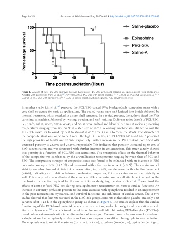
Figure 5. Survival of rats; PEG-20k improves survival duration vs. PEG-20k with saline placebo vs. saline placebo with epinephrine.
†
#
Adapted with permission from Ge et al. [47] . *P = 0.0022 vs. PEG-20k with saline placebo; P = 0.0016 vs. PEG-20k with Saline-A; P =
‡
0.0005 vs. PEG-20k with epinephrine; P = 0.012 vs. saline placebo with epinephrine. PEG: polyethylene glycol
[46]
In another study, Lin et al. prepared the PCL/PEG coated PVA biodegradable composite stents with a
core-shell structure for various applications. The coated yarns were weft knitted into braids followed by
thermal treatment, which resulted in a core-shell structure. In a typical process, the authors fitted the PVA
yarns into a machine, followed by twisting, coating, and weft knitting. Different ratios (wt%) of PCL/PEG,
i.e., 100/0, 90/10, 80/20, 70/30, 60/40, and 50/50 were melted and blended 5 times at various processing
temperatures ranging from 70-100 °C at a step size of 10 °C. A coating machine was utilized to coat the
PCL/PEG mixtures followed by heat treatment at 60 °C for 15 min to form the stents. The diameter of
the composite stent was found to be 3 mm. The high PCL ratios, i.e., PCL/PEG 100:0 and 90:10 possessed
the high porosities of 24.93% and 26.50%, respectively. Further increase in the PEG content from 20-30 wt%
decreased porosity to 23.39% and 23.29%, respectively. This indicated that porosity increased up to 20% of
PEG concentration and was decreased with further increase in concentration. This study clearly showed
that porosity is a function of PCL/PEG concentrations. The synergistic effect on the thermal behavior
of the composite was confirmed by the crystallization temperature ranging between that of PCL and
PEG. The compressive strength of composite stents was found to be enhanced with an increase in PEG
concentration up to 30% (6.15 N) and decreased with a further increment (4.5 N). The maximum cell
viability was also observed at 30% PEG concentration, i.e., > 90%, and decreased with a further increment
(~40%), indicating a correlation between mechanical properties, PEG concentration and cell viability as
well. This study helps to understand the effects of PEG concentration on cell attachment as well as the
[47]
mechanical properties required for the use of PEG for designing the stents. Ge et al. examined the
effects of aortic-infused PEG-20k during cardiopulmonary resuscitation on various cardiac functions. An
increase in coronary perfusion pressure to the same extent as with epinephrine resulted in an improvement
in the post-resuscitation myocardial and cerebral functions and inhibition of cardiac arrest. The in vivo
studies showed that four rats survived in the PEG-20k groups, zero rats in the saline-placebo and only 1 rat
survived after > 24 h in the epinephrine group, as shown in Figure 5. The studies explain that the cardiac
functioning of the PEG-based material depends on its structure, molecular weight and orientation as well.
Similarly, Aykar et al. manufactured the self-standing microfluidic chip using PEG-diacrylate (PEGDA)-
[48]
based hollow microvessels with inner dimensions of 15-73 μm. The macromer solutions were focused onto
a single microchannel hydrodynamically and were subsequently solidified through photopolymerization.
The emphasis was to mimic the arteries (0.1 mm to > 1 cm), arterioles (10-100 μm), capillaries (4-12 μm),