
Page 597 - Read Online
P. 597
Page 6 of 15 Saxena et al. Mini-invasive Surg 2020;4:62 I http://dx.doi.org/10.20517/2574-1225.2020.68
Figure 3. Chemical structure of alginate. G and M refer to α-L-guluronic and β-D-mannuronic residues, respectively. Reprinted (adapted)
with permission from Hecht et al. [38] . Copyright (2016), American Chemical Society
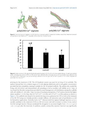
Figure 4. Elastic modulus of 10% alginate dialdehyde-gelatin hydrogels (70-30 wt%) with three angular designs: (1) each layer adhered
to the underlying layer at 90° (0/90°); (2) the second layer adhered to the underlying layer at 45° and the third layer adhered to the
#,
first layer at 135° (0/45/135°); and (3) second layer adhered to the first layer (15°) at 165° (15/165°) ( *P < 0.05). Adapted with
permission from You et al. [22]
printing for the treatment of MI. The 3D bioplotter system was used for printing of the scaffolds. The
authors claimed that 10% oxidation degree of 70 wt% alginate dialdehyde and 30 wt% gelatin concentration
provided the best printability, making it suitable for the 3D printed scaffolds. The authors also seeded the
living cells (EA.hy926) and demonstrated cell spreading as well as excellent cell viability up to 7 days. It
was found that the above proportions provided the most homogenous cell distribution among the scaffolds
with cell viability of > 90% after 7 days. In terms of mechanical properties, 0°/90° pattern showed the higher
elastic modulus (~33 Pa) than that of 0°/45°/135° (~28 kPa) and 15°/165° (~24 kPa) patterns as shown in
Figure 4. The scaffolds were found to be suitable for long-term application for CTE as well as the treatment
of the MI. PCI using such bioinks provide a suitable stiffness, cell interaction and cell proliferation as well
[41]
for biomedical applications. Similarly, Sack et al. demonstrated that alginate hydrogel injections act as a
suitable candidate in the treatment of MI as a left ventricular mid-wall constraint in swine. It was claimed
that the hydrogel injection therapy moderated the elongation in sarcomere lengths from 1.78 ± 0.15 μm to
1.68 ± 0.10 μm after the treatment. In addition, systolic contractility (ejection fraction) was significantly