
Page 577 - Read Online
P. 577
Cossu et al. Mini-invasive Surg 2020;4:60 I http://dx.doi.org/10.20517/2574-1225.2020.52 Page 9 of 15
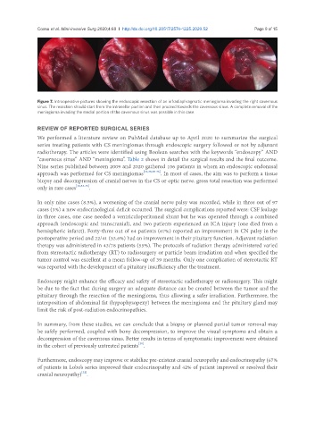
Figure 7. Intraoperative pictures showing the endoscopic resection of an infradiaphragmatic meningioma invading the right cavernous
sinus. The resection should start from the intrasellar portion and then proceed towards the cavernous sinus. A complete removal of the
meningioma invading the medial portion of the cavernous sinus was possible in this case
REVIEW OF REPORTED SURGICAL SERIES
We performed a literature review on PubMed database up to April 2020 to summarize the surgical
series treating patients with CS meningiomas through endoscopic surgery followed or not by adjuvant
radiotherapy. The articles were identified using Boolean searches with the keywords “endoscopy” AND
“cavernous sinus” AND “meningioma”. Table 2 shows in detail the surgical results and the final outcome.
Nine series published between 2009 and 2020 gathered 106 patients in whom an endoscopic endonasal
approach was performed for CS meningiomas [18,28,30-36] . In most of cases, the aim was to perform a tissue
biopsy and decompression of cranial nerves in the CS or optic nerve. gross total resection was performed
only in rare cases [18,33-35] .
In only nine cases (8.5%), a worsening of the cranial nerve palsy was recorded, while in three out of 97
cases (3%) a new endocrinological deficit occurred. The surgical complications reported were: CSF leakage
in three cases, one case needed a ventriculoperitoneal shunt but he was operated through a combined
approach (endoscopic and transcranial), and two patients experienced an ICA injury (one died from a
hemispheric infarct). Forty-three out of 64 patients (67%) reported an improvement in CN palsy in the
postoperative period and 22/41 (53.6%) had an improvement in their pituitary function. Adjuvant radiation
therapy was administered in 43/78 patients (55%). The protocols of radiation therapy administered varied
from stereotactic radiotherapy (RT) to radiosurgery or particle beam irradiation and when specified the
tumor control was excellent at a mean follow-up of 39 months. Only one complication of stereotactic RT
was reported with the development of a pituitary insufficiency after the treatment.
Endoscopy might enhance the efficacy and safety of stereotactic radiotherapy or radiosurgery. This might
be due to the fact that during surgery an adequate distance can be created between the tumor and the
pituitary through the resection of the meningioma, thus allowing a safer irradiation. Furthermore, the
interposition of abdominal fat (hypophysopexy) between the meningioma and the pituitary gland may
limit the risk of post-radiation endocrinopathies.
In summary, from these studies, we can conclude that a biopsy or planned partial tumor removal may
be safely performed, coupled with bony decompression, to improve the visual symptoms and obtain a
decompression of the cavernous sinus. Better results in terms of symptomatic improvement were obtained
[28]
in the cohort of previously untreated patients .
Furthermore, endoscopy may improve or stabilize pre-existent cranial neuropathy and endocrinopathy (67%
of patients in Lobo’s series improved their endocrinopathy and 42% of patient improved or resolved their
[32]
cranial neuropathy) .