
Page 572 - Read Online
P. 572
Page 4 of 15 Cossu et al. Mini-invasive Surg 2020;4:60 I http://dx.doi.org/10.20517/2574-1225.2020.52
Figure 1. Algorithm showing the management of patients with cavernous sinus meningiomas according to the extension of the tumor
and the clinical presentation or the doubt on the histological diagnosis. RTH: radiation therapy
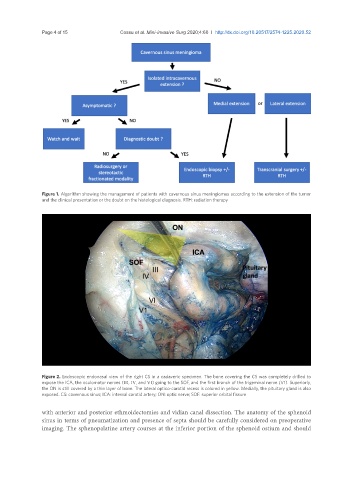
Figure 2. Endoscopic endonasal view of the right CS in a cadaveric specimen. The bone covering the CS was completely drilled to
expose the ICA, the oculomotor nerves (III, IV, and VI) going to the SOF, and the first branch of the trigeminal nerve (V1). Superiorly,
the ON is still covered by a thin layer of bone. The lateral optico-carotid recess is colored in yellow. Medially, the pituitary gland is also
exposed. CS: cavernous sinus; ICA: internal carotid artery; ON: optic nerve; SOF: superior orbital fissure
with anterior and posterior ethmoidectomies and vidian canal dissection. The anatomy of the sphenoid
sinus in terms of pneumatization and presence of septa should be carefully considered on preoperative
imaging. The sphenopalatine artery courses at the inferior portion of the sphenoid ostium and should