
Page 571 - Read Online
P. 571
Cossu et al. Mini-invasive Surg 2020;4:60 I http://dx.doi.org/10.20517/2574-1225.2020.52 Page 3 of 15
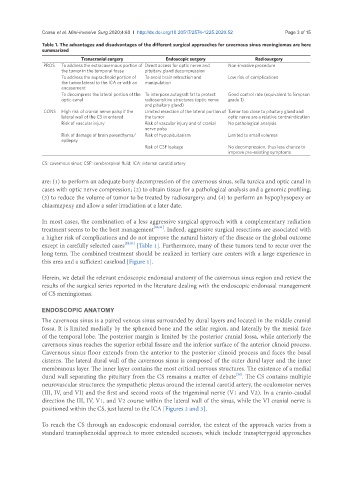
Table 1. The advantages and disadvantages of the different surgical approaches for cavernous sinus meningiomas are here
summarized
Transcranial surgery Endoscopic surgery Radiosurgery
PROS To address the extracavernous portion of Direct access for optic nerve and Non-invasive procedure
the tumor in the temporal fossa pituitary gland decompression
To address the supraclinoid portion of To avoid brain retraction and Low risk of complications
the tumor lateral to the ICA or with an manipulation
encasement
To decompress the lateral portion of the To interpose autograft fat to protect Good control rate (equivalent to Simpson
optic canal radiosensitive structures (optic nerve grade I)
and pituitary gland)
CONS High risk of cranial nerve palsy if the Limited resection of the lateral portion of Tumor too close to pituitary gland and
lateral wall of the CS in entered the tumor optic nerve are a relative contraindication
Risk of vascular injury Risk of vascular injury and of cranial No pathological analysis
nerve palsy
Risk of damage of brain parenchyma/ Risk of hypopituitarism Limited to small volumes
epilepsy
Risk of CSF leakage No decompression, thus less chance to
improve pre-existing symptoms
CS: cavernous sinus; CSF: cerebrospinal fluid; ICA: internal carotid artery
are: (1) to perform an adequate bony decompression of the cavernous sinus, sella turcica and optic canal in
cases with optic nerve compression; (2) to obtain tissue for a pathological analysis and a genomic profiling;
(3) to reduce the volume of tumor to be treated by radiosurgery; and (4) to perform an hypophysopexy or
chiasmapexy and allow a safer irradiation at a later date.
In most cases, the combination of a less aggressive surgical approach with a complementary radiation
treatment seems to be the best management [20,21] . Indeed, aggressive surgical resections are associated with
a higher risk of complications and do not improve the natural history of the disease or the global outcome
except in carefully selected cases [22,23] [Table 1]. Furthermore, many of these tumors tend to recur over the
long term. The combined treatment should be realized in tertiary care centers with a large experience in
this area and a sufficient caseload [Figure 1].
Herein, we detail the relevant endoscopic endonasal anatomy of the cavernous sinus region and review the
results of the surgical series reported in the literature dealing with the endoscopic endonasal management
of CS meningiomas.
ENDOSCOPIC ANATOMY
The cavernous sinus is a paired venous sinus surrounded by dural layers and located in the middle cranial
fossa. It is limited medially by the sphenoid bone and the sellar region, and laterally by the mesial face
of the temporal lobe. The posterior margin is limited by the posterior cranial fossa, while anteriorly the
cavernous sinus reaches the superior orbital fissure and the inferior surface of the anterior clinoid process.
Cavernous sinus floor extends from the anterior to the posterior clinoid process and faces the basal
cisterns. The lateral dural wall of the cavernous sinus is composed of the outer dural layer and the inner
membranous layer. The inner layer contains the most critical nervous structures. The existence of a medial
dural wall separating the pituitary from the CS remains a matter of debate . The CS contains multiple
[24]
neurovascular structures: the sympathetic plexus around the internal carotid artery, the oculomotor nerves
(III, IV, and VI) and the first and second roots of the trigeminal nerve (V1 and V2). In a cranio-caudal
direction the III, IV, V1, and V2 course within the lateral wall of the sinus, while the VI cranial nerve is
positioned within the CS, just lateral to the ICA [Figures 2 and 3].
To reach the CS through an endoscopic endonasal corridor, the extent of the approach varies from a
standard transsphenoidal approach to more extended accesses, which include transpterygoid approaches