
Page 169 - Read Online
P. 169
Crema et al. Minimally invasive esophagectomy in achalasia
ports were positioned in the right hemiclavicular line of the greater curvature. Monopolar electrocauterization
(left hand of the surgeon), 1 cm left from the xiphoid and UltraCision were used for sectioning of the
appendix (aspirator) and 15 cm left from the umbilical short gastric vessels and gastrocolic omentum. The
scar (esophageal separator). gastroepiploic and left gastric vessels were ligated by
double clipping, with preservation of the arch of the
Using a 12-mmHg pneumoperitoneum (CO ), the greater and lesser curvature. No pyloroplasty was
2
procedure was started through ample dissection of the performed during surgical treatment of advanced
esophagogastric transition, restoring the abdominal megaesophagus. After preparing the stomach, the
esophagus with a Penrose drain or a flexible separator cervical esophagus was dissected through a left
(EndoFlex, Medline, Mundelein, IL, USA). Dissection cervicotomy. Owing to the delicate traction of the surgical
was continued with the esophageal body under direct specimen, the esophagus and proximal part of the
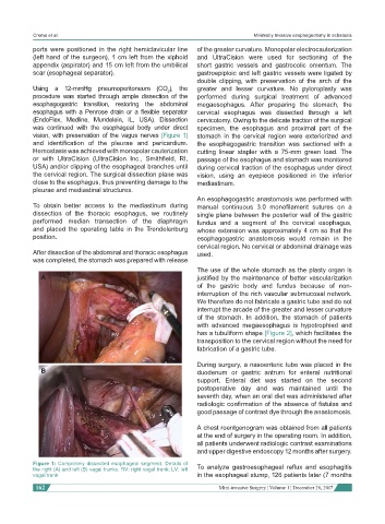
vision, with preservation of the vagus nerves [Figure 1] stomach in the cervical region were exteriorized and
and identification of the pleurae and pericardium. the esophagogastric transition was sectioned with a
Hemostasis was achieved with monopolar cauterization cutting linear stapler with a 75-mm green load. The
or with UltraCision (UltraCision Inc., Smithfield, RI, passage of the esophagus and stomach was monitored
USA) and/or clipping of the esophageal branches until during cervical traction of the esophagus under direct
the cervical region. The surgical dissection plane was vision, using an eyepiece positioned in the inferior
close to the esophagus, thus preventing damage to the mediastinum.
pleurae and mediastinal structures.
An esophagogastric anastomosis was performed with
To obtain better access to the mediastinum during manual continuous 3.0 monofilament sutures on a
dissection of the thoracic esophagus, we routinely single plane between the posterior wall of the gastric
performed median transection of the diaphragm fundus and a segment of the cervical esophagus,
and placed the operating table in the Trendelenburg whose extension was approximately 4 cm so that the
position. esophagogastric anastomosis would remain in the
cervical region. No cervical or abdominal drainage was
After dissection of the abdominal and thoracic esophagus used.
was completed, the stomach was prepared with release
The use of the whole stomach as the plasty organ is
A justified by the maintenance of better vascularization
of the gastric body and fundus because of non-
interruption of the rich vascular submucosal network.
We therefore do not fabricate a gastric tube and do not
interrupt the arcade of the greater and lesser curvature
of the stomach. In addition, the stomach of patients
with advanced megaesophagus is hypotrophied and
RV has a tubuliform shape [Figure 2], which facilitates the
transposition to the cervical region without the need for
fabrication of a gastric tube.
During surgery, a nasoenteric tube was placed in the
B duodenum or gastric antrum for enteral nutritional
support. Enteral diet was started on the second
postoperative day and was maintained until the
seventh day, when an oral diet was administered after
radiologic confirmation of the absence of fistulas and
good passage of contrast dye through the anastomosis.
LV
A chest roentgenogram was obtained from all patients
at the end of surgery in the operating room. In addition,
all patients underwent radiologic contrast examinations
and upper digestive endoscopy 12 months after surgery.
Figure 1: Completely dissected esophageal segment. Details of To analyze gastroesophageal reflux and esophagitis
the right (A) and left (B) vagal trunks. RV: right vagal trunk; LV: left
vagal trunk in the esophageal stump, 126 patients later (7 months
162 Mini-invasive Surgery ¦ Volume 1 ¦ December 28, 2017