
Page 21 - Read Online
P. 21
Gharagozloo et al. Mini-invasive Surg 2020;4:56 I http://dx.doi.org/10.20517/2574-1225.2020.43 Page 7 of 10
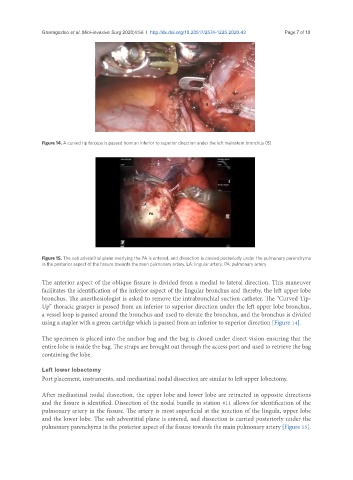
Figure 14. A curved tip forceps is passed from an inferior to superior direction under the left mainstem bronchus (B)
Figure 15. The sub adventitial plane overlying the PA is entered, and dissection is carried posteriorly under the pulmonary parenchyma
in the posterior aspect of the fissure towards the main pulmonary artery. LA: lingular artery; PA: pulmonary artery
The anterior aspect of the oblique fissure is divided from a medial to lateral direction. This maneuver
facilitates the identification of the inferior aspect of the lingular bronchus and thereby, the left upper lobe
bronchus. The anesthesiologist is asked to remove the intrabronchial suction catheter. The “Curved Tip-
Up” thoracic grasper is passed from an inferior to superior direction under the left upper lobe bronchus,
a vessel loop is passed around the bronchus and used to elevate the bronchus, and the bronchus is divided
using a stapler with a green cartridge which is passed from an inferior to superior direction [Figure 14].
The specimen is placed into the anchor bag and the bag is closed under direct vision ensuring that the
entire lobe is inside the bag. The straps are brought out through the access port and used to retrieve the bag
containing the lobe.
Left lower lobectomy
Port placement, instruments, and mediastinal nodal dissection are similar to left upper lobectomy.
After mediastinal nodal dissection, the upper lobe and lower lobe are retracted in opposite directions
and the fissure is identified. Dissection of the nodal bundle in station #11 allows for identification of the
pulmonary artery in the fissure. The artery is most superficial at the junction of the lingula, upper lobe
and the lower lobe. The sub adventitial plane is entered, and dissection is carried posteriorly under the
pulmonary parenchyma in the posterior aspect of the fissure towards the main pulmonary artery [Figure 15].