
Page 16 - Read Online
P. 16
Page 2 of 10 Gharagozloo et al. Mini-invasive Surg 2020;4:56 I http://dx.doi.org/10.20517/2574-1225.2020.43
Figure 1. Port placement for robotic lobectomy of the left chest. AP: assistant port
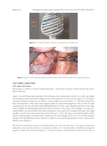
Figure 2. Dissect the inferior pulmonary ligament and remove station #9 and #8 nodes. IPL: inferior pulmonary ligament
LEFT SIDED LOBECTOMY
Left upper lobectomy
Instruments: 0° and/or 30° down viewing endoscope, 5 mm thoracic grasper, Cadiere forceps and curved
bipolar dissector.
Figure 1 shows left sided port placement. The technique of port placement is similar to the right side. Begin
by dividing the inferior pulmonary ligament and removing station #9 and #8 nodes [Figure 2]. The lung is
retracted medially and anteriorly in order to remove lymph nodes from station #7. After the stomach has
been decompressed, at this stage, some surgeons prefer to remove the nasogastric tube in order to create
a greater space for the subcarinal and mediastinal dissection. Next, open the pleura anterior to the vagus
nerve. Identify the left mainstem bronchus and stay inferior to the edge of the cartilage. The station #7
nodal bundle is accessed between the inferior pulmonary vein and the left mainstem bronchus. The nodal
bundle is traced to the carina and is then removed [Figure 3]. Next, the lung is retracted inferiorly, and the
pleura overlying station #5 nodal bundle is opened in the lower margin of the aortic arch and the superior
margin of the left pulmonary artery. Station #5 nodes are removed paying attention to the location of the
phrenic nerve [Figure 4].
The left main pulmonary artery is identified above the left main bronchus. The space between the
pulmonary artery and the bronchus is opened and station #10L nodal bundle is identified overlying the
superior border of the bronchus [Figure 5]. The space between the pulmonary artery and the aorta is