
Page 19 - Read Online
P. 19
Gharagozloo et al. Mini-invasive Surg 2020;4:56 I http://dx.doi.org/10.20517/2574-1225.2020.43 Page 5 of 10
Figure 9. In the main fissure, the sub adventitial plane over the PA is entered, and dissection is carried posteriorly under the pulmonary
parenchyma in the posterior aspect of the fissure towards the main PA. PA: pulmonary artery
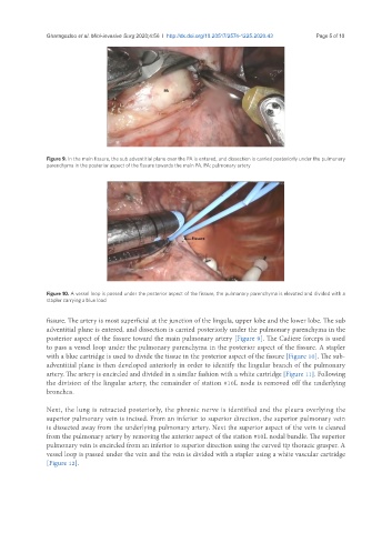
Figure 10. A vessel loop is passed under the posterior aspect of the fissure, the pulmonary parenchyma is elevated and divided with a
stapler carrying a blue load
fissure. The artery is most superficial at the junction of the lingula, upper lobe and the lower lobe. The sub
adventitial plane is entered, and dissection is carried posteriorly under the pulmonary parenchyma in the
posterior aspect of the fissure toward the main pulmonary artery [Figure 9]. The Cadiere forceps is used
to pass a vessel loop under the pulmonary parenchyma in the posterior aspect of the fissure. A stapler
with a blue cartridge is used to divide the tissue in the posterior aspect of the fissure [Figure 10]. The sub-
adventitial plane is then developed anteriorly in order to identify the lingular branch of the pulmonary
artery. The artery is encircled and divided in a similar fashion with a white cartridge [Figure 11]. Following
the division of the lingular artery, the remainder of station #10L node is removed off the underlying
bronchus.
Next, the lung is retracted posteriorly, the phrenic nerve is identified and the pleura overlying the
superior pulmonary vein is incised. From an inferior to superior direction, the superior pulmonary vein
is dissected away from the underlying pulmonary artery. Next the superior aspect of the vein is cleared
from the pulmonary artery by removing the anterior aspect of the station #10L nodal bundle. The superior
pulmonary vein is encircled from an inferior to superior direction using the curved tip thoracic grasper. A
vessel loop is passed under the vein and the vein is divided with a stapler using a white vascular cartridge
[Figure 12].