
Page 18 - Read Online
P. 18
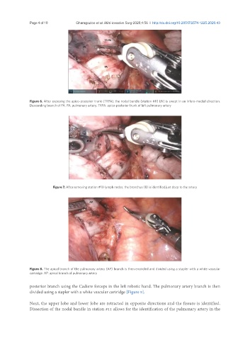
Page 4 of 10                               Gharagozloo et al. Mini-invasive Surg 2020;4:56  I  http://dx.doi.org/10.20517/2574-1225.2020.43


               Figure 6. After exposing the apico-posterior trunk (TRPA), the nodal bundle (station #10 LN) is swept in an infero-medial direction.
               Descending branch of PA. PA: pulmonary artery; TRPA: apico-posterior trunk of left pulmonary artery


                          Figure 7. After removing station #10 lymph nodes, the bronchus (B) is identified just deep to the artery


               Figure 8. The apical branch of the pulmonary artery (AP) branch is then encircled and divided using a stapler with a white vascular
               cartridge. AP: apical branch of pulmonary artery


               posterior branch using the Cadiere forceps in the left robotic hand. The pulmonary artery branch is then
               divided using a stapler with a white vascular cartridge [Figure 8].

               Next, the upper lobe and lower lobe are retracted in opposite directions and the fissure is identified.
               Dissection of the nodal bundle in station #11 allows for the identification of the pulmonary artery in the