
Page 17 - Read Online
P. 17
Gharagozloo et al. Mini-invasive Surg 2020;4:56 I http://dx.doi.org/10.20517/2574-1225.2020.43 Page 3 of 10
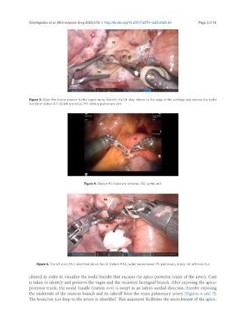
Figure 3. Open the pleura anterior to the vagus nerve. Identify the LB, stay inferior to the edge of the cartilage and remove the nodal
bundle of station #7. LB: left bronchus; IPV: inferior pulmonary vein
Figure 4. Station #5 nodes are removed. AO: aortic arch
Figure 5. The left main PA is identified above the LB. Station #10L nodes are removed. PA: pulmonary artery; LB: left bronchus
cleared in order to visualize the nodal bundle that encases the apico-posterior trunk of the artery. Care
is taken to identify and preserve the vagus and the recurrent laryngeal branch. After exposing the apico-
posterior trunk, the nodal bundle (station #10) is swept in an infero-medial direction, thereby exposing
the underside of the truncus branch and its takeoff from the main pulmonary artery [Figures 6 and 7].
The bronchus just deep to the artery is identified. This maneuver facilitates the encirclement of the apico-