
Page 17 - Read Online
P. 17
Page 4 of 10 Tokairin et al. Mini-invasive Surg 2020;4:32 I http://dx.doi.org/10.20517/2574-1225.2020.23
AA B
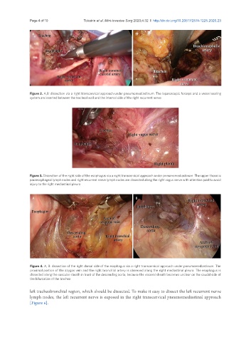
Figure 2. A,B: dissection via a right transcervical approach under pneumomediastinum. The laparoscopic forceps and a vessel sealing
system are inserted between the tracheal wall and the internal side of the right recurrent nerve
Figure 3. Dissection of the right side of the esophagus via a right transcervical approach under pneumomediastinum. The upper thoracic
paraesophageal lymph nodes and right recurrent nerve lymph nodes are dissected along the right vagus nerve with attention paid to avoid
injury to the right mediastinal pleura
A B
Figure 4. A, B: dissection of the right dorsal side of the esophagus via a right transcervical approach under pneumomediastinum. The
proximal portion of the azygos vein and the right bronchial artery is observed along the right mediastinal pleura. The esophagus is
dissected along the vascular sheath in front of the descending aorta, because the visceral sheath becomes unclear on the caudal side of
the bifurcation of the trachea
left tracheobronchial region, which should be dissected. To make it easy to dissect the left recurrent nerve
lymph nodes, the left recurrent nerve is exposed in the right transcervical pneumomediastinal approach
[Figure 6].