
Page 16 - Read Online
P. 16
Tokairin et al. Mini-invasive Surg 2020;4:32 I http://dx.doi.org/10.20517/2574-1225.2020.23 Page 3 of 10
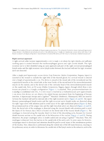
Figure 1. The location of the ports and single-port laparoscopic access devices. This schematic illustration shows single-port laparoscopic
access devices in the bilateral cervical area and ports in the abdominal area. A single-port laparoscopic access device with three 5-mm
trocars is placed in a triangle configuration. Approximately 3 cm above this device, a 5-mm trocar is inserted as a mediastinoscopic port
Right cervical approach
A right cervical collar incision (approximately 6 cm) is made 3 cm above the right clavicle, and sufficient
working space is created between the tracheoesophageal groove and right carotid sheath. The right
recurrent nerve is first identified using an open approach and part of the right cervical paraesophageal
lymph nodes and the right recurrent nerve lymph nodes between the tracheal wall and the right recurrent
nerve are dissected.
After a single-port laparoscopic access device (Lap Protector; Hakko Corporation, Nagano, Japan) is
inserted in the wound to exclude the right lobe of the thyroid gland, the cervical wound is sutured
over a length of approximately 4 cm. The device is placed at the dorsal side of the sternohyoid and the
scapulohyoid muscles at the cranial side, at the inner border of the sternal head of the sternocleidomastoid
muscle on the outside, and at the dorsal side of the clavicular head of the sternocleidomastoid muscle
on the caudal side. Next, an EZ access (Hakko Corporation, Nagano, Japan), through which three 5-mm
trocars are placed in a triangle configuration [Figure 1], is attached. Then, pneumomediastinum (to
8 mmHg) is established with CO . A 5-mm trocar is inserted as a mediastinoscopic port approximately
2
3 cm above this device; we can observe the whole forceps movement from the beginning of forceps
TM
insertion. A laparoscopic forceps and a LigaSure Maryland (Medtronic, Minneapolis, MN) are inserted
between the tracheal wall and the internal side of the right recurrent nerve [Figure 2A and B]. The upper
thoracic paraesophageal lymph nodes and the right recurrent nerve lymph nodes are dissected along
the right vagus nerve with attention paid to avoid injury of the right mediastinal pleura [Figure 3]; then,
the proximal portion of the azygos vein and the right bronchial artery are dissected [Figure 4A and B].
Next, the dorsal side of the esophagus is dissected along the visceral sheath with attention paid to avoid
the injury of the thoracic duct, which is located on the dorsal side of the visceral sheath [Figure 5]. The
esophagus is dissected along the vascular sheath in front of the descending aorta, because the visceral
sheath becomes unclear on the caudal side of the bifurcation of the trachea [Figure 4 A and B]. During
TM
dissection, the proper esophageal artery is double sealed and cut using a LigaSure Maryland. Then, the
esophagus is dissected from the trachea on the caudal side of the aortic arch. In some cases, the broncho-
oesophagus muscle is developed and attention should be paid to avoid injury of the membranous portion
of the trachea. Next, dissection is performed along the ventral side of the left main bronchus, reaching to
the pulmonary artery, in order to determine the caudal side of the lymph nodes in the subaortic arch to the