
Page 18 - Read Online
P. 18
Tokairin et al. Mini-invasive Surg 2020;4:32 I http://dx.doi.org/10.20517/2574-1225.2020.23 Page 5 of 10
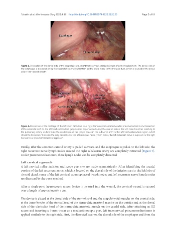
Figure 5. Dissection of the dorsal side of the esophagus via a right transcervical approach under pneumomediastinum. The dorsal side of
the esophagus is dissected along the visceral sheath with attention paid to avoid injury to the thoracic duct, which is located on the dorsal
side of the visceral sheath
Figure 6. Dissection of the cartilage of the left main bronchus via a right transcervical approach under pneumomediastinum. Dissection
of the subaortic arch to the left tracheobronchial lymph nodes is performed along the ventral side of the left main bronchus reaching to
the pulmonary artery to determine the caudal side of the lymph nodes in the subaortic arch to the left tracheobronchial region, which
should be dissected. To enable the easy dissection of the left recurrent nerve lymph nodes, the left recurrent nerve is exposed via the right
transcervical pneumomediastinal approach
Finally, after the common carotid artery is pulled outward and the esophagus is pulled to the left side, the
right recurrent nerve lymph nodes around the right subclavian artery are completely retrieved [Figure 7].
Under pneumomediastinum, these lymph nodes can be completely dissected.
Left cervical approach
A left cervical collar incision and scope port site are made symmetrically. After identifying the cranial
portion of the left recurrent nerve, which is located on the dorsal side of the inferior part in the left lobe of
thyroid gland, some of the left cervical paraesophageal lymph nodes and left recurrent nerve lymph nodes
are dissected by the open method.
After a single-port laparoscopic access device is inserted into the wound, the cervical wound is sutured
over a length of approximately 4 cm.
The device is placed at the dorsal side of the sternohyoid and the scapulohyoid muscles on the cranial side,
at the inner border of the sternal head of the sternocleidomastoid muscle on the outside and at the dorsal
side of the clavicular head of the sternocleidomastoid muscle on the caudal side. After attaching an EZ
access and inserting a 5-mm trocar as a mediastinoscopic port, left transcervical pneumomediastinum is
applied similarly to the right side. First, the dissected layer on the dorsal side of the esophagus and from the