
Page 20 - Read Online
P. 20
Tokairin et al. Mini-invasive Surg 2020;4:32 I http://dx.doi.org/10.20517/2574-1225.2020.23 Page 7 of 10
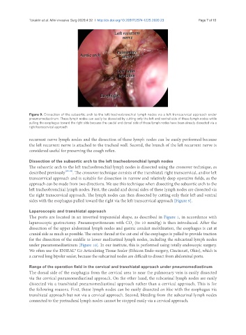
Figure 9. Dissection of the subaortic arch to the left tracheobronchial lymph nodes via a left transcervical approach under
pneumomediastinum. These lymph nodes can easily be dissected by cutting only the left and ventral side of these lymph nodes while
pulling the esophagus toward the right side because the caudal and dorsal side of these lymph nodes have been already dissected via a
right transcervical approach
recurrent nerve lymph nodes and the dissection of these lymph nodes can be easily performed because
the left recurrent nerve is attached to the tracheal wall. Second, the branch of the left recurrent nerve is
considered useful for preserving the cough reflex.
Dissection of the subaortic arch to the left tracheobronchial lymph nodes
The subaortic arch to the left tracheobronchial lymph nodes is dissected using the crossover technique, as
described previously [15-18] . The crossover technique consists of the transhiatal, right transcervical, and/or left
transcervical approach and is suitable for dissection in narrow and relatively deep operative fields, as the
approach can be made from two directions. We use this technique when dissecting the subaortic arch to the
left tracheobronchial lymph nodes. First, the caudal and dorsal sides of these lymph nodes are dissected via
the right transcervical approach. The lymph nodes can then dissected by cutting only their left and ventral
sides with the esophagus pulled toward the right via the left transcervical approach [Figure 9].
Laparoscopic and transhiatal approach
The ports are located in an inverted trapezoidal shape, as described in Figure 1, in accordance with
laparoscopic gastrectomy. Pneumoperitoneum with CO (to 10 mmHg) is then introduced. After the
2
dissection of the upper abdominal lymph nodes and gastric conduit mobilization, the esophagus is cut at
cranial side as much as possible. The suture thread at the cut end of the esophagus is pulled to provide traction
for the dissection of the middle to lower mediastinal lymph nodes, including the subcarinal lymph nodes
under pneumomediastinum [Figure 10]. In our institute, this is performed using totally endoscopic surgery.
We often use the ENSEAL® G2 Articulating Tissue Sealer (Ethicon Endo-surgery, Cincinnati, Ohio), which is
a curved long bipolar sealer, because the subcarinal nodes are difficult to dissect from abdominal ports.
Range of the operation field in the cervical and transhiatal approach under pneumomediastinum
The dorsal side of the esophagus from the cervical area to near the pulmonary vein is easily dissected
via the cervical pneumomediastinal approach. On the other hand, the subcarinal lymph nodes are easily
dissected via a transhiatal pneumomediastinal approach rather than a cervical approach. This is for
the following reasons. First, these lymph nodes can be easily dissected en bloc with the esophagus via
transhiatal approach but not via a cervical approach. Second, bleeding from the subcarinal lymph nodes
connected to the pretracheal lymph nodes cannot be stopped easily via a cervical approach.