
Page 37 - Read Online
P. 37
Page 6 of 12 Tschuor et al. Mini-invasive Surg 2020;4:72 I http://dx.doi.org/10.20517/2574-1225.2020.39
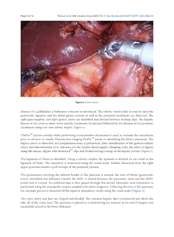
Figure 4. Kocherization
absence of a gallbladder a Nathanson retractor is introduced. The robotic vessel sealer is used to open the
gastrocolic ligament and the distal gastric antrum as well as the proximal duodenum are dissected. The
right gastroepiploic and right gastric artery are identified and divided between locking clips. The hepatic
flexure of the colon is taken down and the duodenum Kocherized followed by the division of the proximal
duodenum using a 60-mm robotic stapler [Figure 4].
TilePro TM picture overlay while performing intraoperative ultrasound is used to evaluate the vasculature
TM
prior to division of vessels. Fluorescence imaging FireFly assists in identifying the biliary structures. The
hepatic artery is dissected, and lymphadenectomy is performed. After identification of the gastroduodenal
artery and determination of its relevance for the hepatic blood supply (clamping trial), the artery is ligated
TM
using silk sutures, clipped with Hemolock clips and divided leaving a stump on the hepatic portion [Figure 5].
The ligament of Treitz is identified. Using a robotic stapler, the jejunum is divided 20 cm distal to the
ligament of Treitz. The mesentery is transected using the vessel sealer. Further dissection from the right
upper quadrant enables a pull through of the proximal jejunum.
The peritoneum overlying the inferior border of the pancreas is incised, the vein of Henle (gastrocolic
trunc) identified and followed towards the SMV. A tunnel between the pancreatic neck and the SMV/
portal vein is created. An umbilical tape is then passed through this tunnel. Pancreatic neck transection is
performed using the monopolar scissors coupled with saline irrigation. Following division of the pancreas,
the uncinate process is dissected off the superior mesenteric vessels using the vessel sealer [Figure 6].
The cystic artery and duct are clipped and divided. The common hepatic duct is transected just above the
take off of the cystic duct. The specimen is placed in a retrieval bag for removal at the end of surgery and
meanwhile placed in the lower abdomen.