
Page 30 - Read Online
P. 30
Page 8 of 15 Kumar et al. Mini-invasive Surg 2018;2:19 I http://dx.doi.org/10.20517/2574-1225.2018.26
Pubic symphysis
Obturator nerve
Left external iliac vessels Paravesical space
Pelvic floor
Internal iliac artery
Rectum
Left ureter
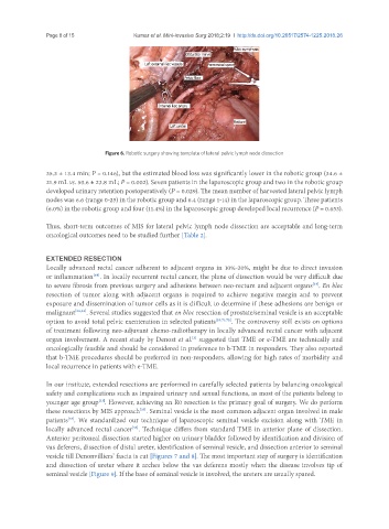
Figure 6. Robotic surgery showing template of lateral pelvic lymph node dissection
35.3 ± 13.4 min; P = 0.146), but the estimated blood loss was significantly lower in the robotic group (34.6 ±
21.9 mL vs. 50.6 ± 23.8 mL; P = 0.002). Seven patients in the laparoscopic group and two in the robotic group
developed urinary retention postoperatively (P = 0.029). The mean number of harvested lateral pelvic lymph
nodes was 6.6 (range 0-25) in the robotic group and 6.4 (range 1-14) in the laparoscopic group. Three patients
(6.0%) in the robotic group and four (11.4%) in the laparoscopic group developed local recurrence (P = 0.653).
Thus, short-term outcomes of MIS for lateral pelvic lymph node dissection are acceptable and long-term
oncological outcomes need to be studied further [Table 2].
EXTENDED RESECTION
Locally advanced rectal cancer adherent to adjacent organs in 10%-20%, might be due to direct invasion
or inflammation . In locally recurrent rectal cancer, the plane of dissection would be very difficult due
[68]
to severe fibrosis from previous surgery and adhesions between neo-rectum and adjacent organs . En bloc
[24]
resection of tumor along with adjacent organs is required to achieve negative margin and to prevent
exposure and dissemination of tumor cells as it is difficult to determine if these adhesions are benign or
malignant [23,33] . Several studies suggested that en bloc resection of prostate/seminal vesicle is an acceptable
option to avoid total pelvic exenteration in selected patients [23,71,72] . The controversy still exists on options
of treatment following neo-adjuvant chemo-radiotherapy in locally advanced rectal cancer with adjacent
organ involvement. A recent study by Denost et al. suggested that TME or e-TME are technically and
[5]
oncologically feasible and should be considered in preference to b-TME in responders. They also reported
that b-TME procedures should be preferred in non-responders, allowing for high rates of morbidity and
local recurrence in patients with e-TME.
In our institute, extended resections are performed in carefully selected patients by balancing oncological
safety and complications such as impaired urinary and sexual functions, as most of the patients belong to
[33]
younger age group . However, achieving an R0 resection is the primary goal of surgery. We do perform
these resections by MIS approach . Seminal vesicle is the most common adjacent organ involved in male
[35]
patients . We standardized our technique of laparoscopic seminal vesicle excision along with TME in
[73]
locally advanced rectal cancer . Technique differs from standard TME in anterior plane of dissection.
[35]
Anterior peritoneal dissection started higher on urinary bladder followed by identification and division of
vas deferens, dissection of distal ureter, identification of seminal vesicle, and dissection anterior to seminal
vesicle till Denonvilliers’ fascia is cut [Figures 7 and 8]. The most important step of surgery is identification
and dissection of ureter where it arches below the vas deferens mostly when the disease involves tip of
seminal vesicle [Figure 9]. If the base of seminal vesicle is involved, the ureters are usually spared.