
Page 26 - Read Online
P. 26
Page 4 of 15 Kumar et al. Mini-invasive Surg 2018;2:19 I http://dx.doi.org/10.20517/2574-1225.2018.26
Pubic symphysis
Dorsal venous complex
Bladder
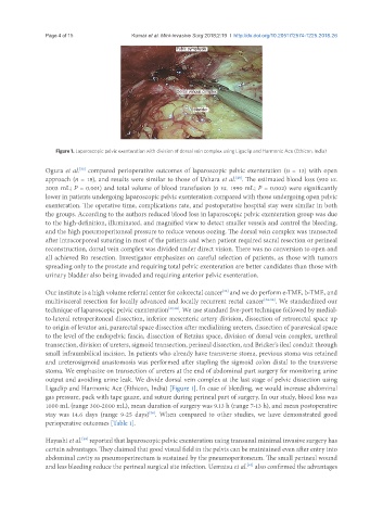
Figure 1. Laparoscopic pelvic exenteration with division of dorsal vein complex using Ligaclip and Harmonic Ace (Ethicon, India)
Ogura et al. compared perioperative outcomes of laparoscopic pelvic exenteration (n = 13) with open
[32]
approach (n = 18), and results were similar to those of Uehara et al. . The estimated blood loss (930 vs.
[29]
3003 mL; P = 0.001) and total volume of blood transfusion (0 vs. 1990 mL; P = 0.002) were significantly
lower in patients undergoing laparoscopic pelvic exenteration compared with those undergoing open pelvic
exenteration. The operative time, complications rate, and postoperative hospital stay were similar in both
the groups. According to the authors reduced blood loss in laparoscopic pelvic exenteration group was due
to the high-definition, illuminated, and magnified view to detect smaller vessels and control the bleeding,
and the high pneumoperitoneal pressure to reduce venous oozing. The dorsal vein complex was transected
after intracorporeal suturing in most of the patients and when patient required sacral resection or perineal
reconstruction, dorsal vein complex was divided under direct vision. There was no conversion to open and
all achieved R0 resection. Investigator emphasizes on careful selection of patients, as those with tumors
spreading only to the prostate and requiring total pelvic exenteration are better candidates than those with
urinary bladder also being invaded and requiring anterior pelvic exenteration.
Our institute is a high volume referral center for colorectal cancer and we do perform e-TME, b-TME, and
[33]
multivisceral resection for locally advanced and locally recurrent rectal cancer [34-36] . We standardized our
technique of laparoscopic pelvic exenteration [37,38] . We use standard five-port technique followed by medial-
to-lateral retroperitoneal dissection, inferior mesenteric artery division, dissection of retrorectal space up
to origin of levator ani, pararectal space dissection after medializing ureters, dissection of paravesical space
to the level of the endopelvic fascia, dissection of Retzius space, division of dorsal vein complex, urethral
transection, division of ureters, sigmoid transection, perineal dissection, and Bricker’s ileal conduit through
small infraumbilical incision. In patients who already have transverse stoma, previous stoma was retained
and ureterosigmoid anastomosis was performed after stapling the sigmoid colon distal to the transverse
stoma. We emphasize on transection of ureters at the end of abdominal part surgery for monitoring urine
output and avoiding urine leak. We divide dorsal vein complex at the last stage of pelvic dissection using
Ligaclip and Harmonic Ace (Ethicon, India) [Figure 1]. In case of bleeding, we would increase abdominal
gas pressure, pack with tape gauze, and suture during perineal part of surgery. In our study, blood loss was
1000 mL (range 300-2000 mL), mean duration of surgery was 9.13 h (range 7-13 h), and mean postoperative
stay was 14.6 days (range 9-25 days) . When compared to other studies, we have demonstrated good
[38]
perioperative outcomes [Table 1].
Hayashi et al. reported that laparoscopic pelvic exenteration using transanal minimal invasive surgery has
[39]
certain advantages. They claimed that good visual field in the pelvis can be maintained even after entry into
abdominal cavity as pneumoperirectum is sustained by the pneumoperitoneum. The small perineal wound
and less bleeding reduce the perineal surgical site infection. Uematsu et al. also confirmed the advantages
[40]