
Page 75 - Read Online
P. 75
Zhu et al. Mini-invasive Surg 2023;7:12 https://dx.doi.org/10.20517/2574-1225.2022.117 Page 9 of 13
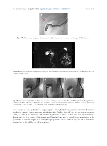
Figure 13. (A) lumen-apposing metal stent between the gallbladder and duodenum was seen during an ERCP (red arrow).
Figure 14. Magnetic resonance cholangiopancreatography (MRCP) with secretin and abnormal tethering of D1 to the gallbladder was
observed (yellow arrow).
Figure 15. ERCP shows a patent fistula. (A) balloon occlusion cholangiogram of the CBD with contrast filling the cystic duct, gallbladder
then entering the duodenum via the fistula (red circle) and (B) a wire passed retrograde through the fistula into the gallbladder
demonstrates a patent fistula; (C) the ERCP patent fistula closed with a Steris Padlock clip.
There was no air in the gallbladder to suggest a patent fistula, but following a multidisciplinary discussion,
we planned an ERCP to attempt endoscopic closure of the fistula if detected, prior to operative intervention.
During the ERCP, the duodenal bulb was investigated and there was a clear persistent fistula with bile
flowing into the first portion of the duodenum [Figure 15]. A wire was passed through the fistula to the
gallbladder to confirm its presence. Using twin graspers and a Steris Padlock clip, the fistula was closed
using a piece of the gallbladder wall as a buttress.