
Page 74 - Read Online
P. 74
Page 8 of 13 Zhu et al. Mini-invasive Surg 2023;7:12 https://dx.doi.org/10.20517/2574-1225.2022.117
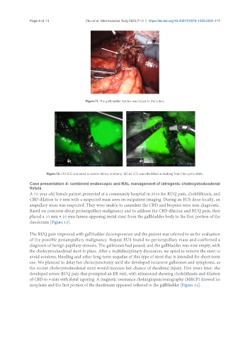
Figure 11. The gallbladder fundus was fused to the colon.
Figure 12. (A) ICG was used to assess biliary anatomy; (B) no ICG was identified as leaking from the cystic plate.
Case presentation 4: combined endoscopic and RAL management of iatrogenic cholecystoduodenal
fistula
A 55-year-old female patient presented at a community hospital in 2016 for RUQ pain, cholelithiasis, and
CBD dilation to 9 mm with a suspected mass seen on outpatient imaging. During an EUS done locally, an
ampullary mass was suspected. They were unable to cannulate the CBD and biopsies were non-diagnostic.
Based on concerns about periampullary malignancy and to address the CBD dilation and RUQ pain, they
placed a 10 mm × 10 mm lumen-apposing metal stent from the gallbladder body to the first portion of the
duodenum [Figure 13].
The RUQ pain improved with gallbladder decompression and the patient was referred to us for evaluation
of the possible periampullary malignancy. Repeat EUS found no periampullary mass and confirmed a
diagnosis of benign papillary stenosis. The gallstones had passed, and the gallbladder was now empty, with
the cholecystoduodenal stent in place. After a multidisciplinary discussion, we opted to remove the stent to
avoid erosions, bleeding and other long-term sequelae of this type of stent that is intended for short-term
use. We planned to delay her cholecystectomy until she developed recurrent gallstones and symptoms, as
the recent cholecystoduodenal stent would increase her chance of duodenal injury. Five years later, she
developed severe RUQ pain that prompted an ER visit, with ultrasound showing cholelithiasis and dilation
of CBD to 9 mm with distal tapering. A magnetic resonance cholangiopancreatography (MRCP) showed no
neoplasm and the first portion of the duodenum appeared tethered to the gallbladder [Figure 14].