
Page 73 - Read Online
P. 73
Zhu et al. Mini-invasive Surg 2023;7:12 https://dx.doi.org/10.20517/2574-1225.2022.117 Page 7 of 13
Figure 9. (A) Gallstones in the gallbladder and common bile duct and a stent are observed on a radiograph; (B) a cholecystocolonic
fistula is observed (white arrow) on a radiograph.
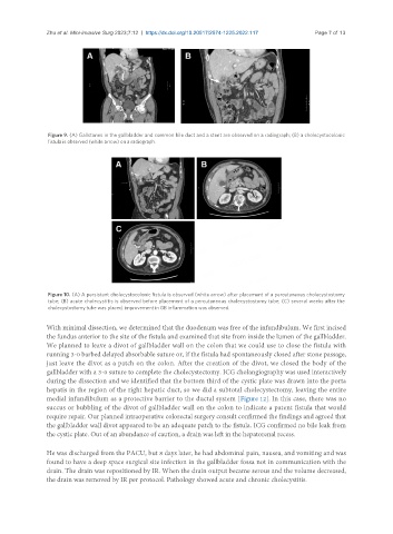
Figure 10. (A) A persistent cholecystocolonic fistula is observed (white arrow) after placement of a percutaneous cholecystostomy
tube; (B) acute cholecystitis is observed before placement of a percutaneous cholecystostomy tube; (C) several weeks after the
cholecystostomy tube was placed, improvement in GB inflammation was observed.
With minimal dissection, we determined that the duodenum was free of the infundibulum. We first incised
the fundus anterior to the site of the fistula and examined that site from inside the lumen of the gallbladder.
We planned to leave a divot of gallbladder wall on the colon that we could use to close the fistula with
running 3-0 barbed delayed absorbable suture or, if the fistula had spontaneously closed after stone passage,
just leave the divot as a patch on the colon. After the creation of the divot, we closed the body of the
gallbladder with a 3-0 suture to complete the cholecystectomy. ICG cholangiography was used interactively
during the dissection and we identified that the bottom third of the cystic plate was drawn into the porta
hepatis in the region of the right hepatic duct, so we did a subtotal cholecystectomy, leaving the entire
medial infundibulum as a protective barrier to the ductal system [Figure 12]. In this case, there was no
succus or bubbling of the divot of gallbladder wall on the colon to indicate a patent fistula that would
require repair. Our planned intraoperative colorectal surgery consult confirmed the findings and agreed that
the gallbladder wall divot appeared to be an adequate patch to the fistula. ICG confirmed no bile leak from
the cystic plate. Out of an abundance of caution, a drain was left in the hepatorenal recess.
He was discharged from the PACU, but 8 days later, he had abdominal pain, nausea, and vomiting and was
found to have a deep space surgical site infection in the gallbladder fossa not in communication with the
drain. The drain was repositioned by IR. When the drain output became serous and the volume decreased,
the drain was removed by IR per protocol. Pathology showed acute and chronic cholecystitis.