
Page 72 - Read Online
P. 72
Page 6 of 13 Zhu et al. Mini-invasive Surg 2023;7:12 https://dx.doi.org/10.20517/2574-1225.2022.117
Figure 7. “White” bile was observed in the cystic duct orifice reflecting chronic obstruction of the cystic duct, in this case by a biliary
stent and a large cholesterol gallstone.
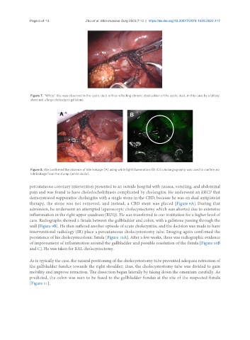
Figure 8. We confirmed the absence of bile leakage (A) using white light illumination (B) ICG cholangiography was used to confirm no
bile leakage from the stump (white circle).
percutaneous coronary intervention presented to an outside hospital with nausea, vomiting, and abdominal
pain and was found to have choledocholithiasis complicated by cholangitis. He underwent an ERCP that
demonstrated suppurative cholangitis with a single stone in the CBD; because he was on dual antiplatelet
therapy, the stone was not removed, and instead, a CBD stent was placed [Figure 9A]. During that
admission, he underwent an attempted laparoscopic cholecystectomy which was aborted due to extensive
inflammation in the right upper quadrant (RUQ). He was transferred to our institution for a higher level of
care. Radiographs showed a fistula between the gallbladder and colon, with a gallstone passing through the
wall [Figure 9B]. He then suffered another episode of acute cholecystitis, and the decision was made to have
interventional radiology (IR) place a percutaneous cholecystostomy tube. Imaging again confirmed the
persistence of his cholecystocolonic fistula [Figure 10A]. After a few weeks, there was radiographic evidence
of improvement of inflammation around the gallbladder and possible resolution of the fistula [Figure 10B
and C]. He was taken for RAL cholecystectomy.
As is typically the case, the natural positioning of the cholecystostomy tube prevented adequate retraction of
the gallbladder fundus towards the right shoulder; thus, the cholecystostomy tube was divided to gain
mobility and improve retraction. The dissection began laterally by taking down the omentum carefully. As
predicted, the colon was seen to be fused to the gallbladder fundus at the site of the suspected fistula
[Figure 11].