
Page 50 - Read Online
P. 50
Kitahama et al. PLPED with EMG monitoring under general anesthesia
METHODS calculated from CT and plain roentgenography (both
anteroposterior and lateral views) [Figure 2B and C].
Ethics and patient consent The calculated points on were drawn on patient’s skin
This study was approved by the Ethics committee along with the anatomical landmarks (vertebral body,
of the Omaezaki Municipal Hospital and all involved spinous process, transverse process, and iliac crest)
patients gave consent. to avoid incorrect puncture [Figure 3].
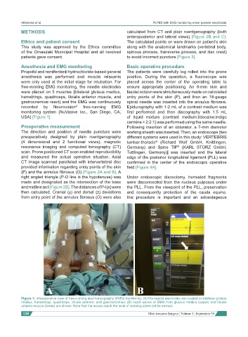
Anesthesia and EMG monitoring Basic operative procedure
Propofol and remifentanil hydrochloride-based general The patients were carefully log rolled into the prone
anesthesia was performed and muscle relaxants position. During the operation, a fluoroscope was
were only used at the initial stage for intubation. For placed across the center of the operating table to
free-running EMG monitoring, the needle electrodes ensure appropriate positioning. An 8-mm skin and
were placed on 5 muscles (bilateral gluteus medius, fascia incision were simultaneously made on calculated
hamstrings, quadriceps, tibialis anterior muscle, and entry points of the skin (P), and then an 18-gauge
gastrocnemius react) and the EMG was continuously spinal needle was inserted into the annulus fibrosus.
recorded by Neurovision ® free-running EMG Epidurography with 1-2 mL of a contrast medium was
monitoring system (NuVasive Inc., San Diego, CA, first performed and then discography with 1.5 mL
USA) [Figure 1]. of liquid mixture (contrast medium:lidocaine:indigo
carmine = 2:2:1) was performed using the same needle.
Preoperative measurement Following insertion of an obturator, a 7-mm diameter
The direction and position of needle puncture were working sheath was inserted. Then, an endoscope [two
preoperatively designed by plain roentgenography different systems were used in this study: VERTEBRIS
(4 dimensional and 2 functional views), magnetic lumbar-thoracic (Richard Wolf GmbH, Knittlingen,
®
resonance imaging and computed tomography (CT) Germany) and Spine TIP (KARL STORZ GmbH,
®
scan. Prone positioned CT scan enabled reproducibility Tuttlingen, Germany)] was inserted and the lateral
and measured the actual operative situation. Axial edge of the posterior longitudinal ligament (PLL) was
CT image scanned paralleled with intervertebral disc confirmed in the center of the endoscopic operative
provided information regarding entry points of the skin field [Figure 4A].
(P) and the annulus fibrosus (O) [Figure 2A and B]. A
right angled triangle (P-O line is the hypotenuse) was Under endoscopic discectomy, herniated fragments
made and designated as the intersection of the base were disconnected from the nucleus pulposus under
and midline as I [Figure 2B]. The distances of P-I(x) were the PLL. From the viewpoint of the PLL, preservation
then calculated. Cranial (y) and dorsal (z) deviations and consequently protection of the cauda equine,
from entry point of the annulus fibrosus (O) were also this procedure is important and an advantageous
Figure 1: Intraoperative view of free-running electromyography (EMG) monitoring. (A)The needle electrodes are located on bilateral gluteus
medius, hamstrings, quadriceps, tibialis anterior, and gastrocnemius; (B) exact waves of EMG from gluteus medius (upper) and tibialis
anterior muscle (lower) are shown. Note that the waves reach the level of warning alarm (white arrows)
110 Mini-invasive Surgery ¦ Volume 1 ¦ September 30