
Page 47 - Read Online
P. 47
Page 8 of 14 Farinha et al. Mini-invasive Surg 2023;7:38 https://dx.doi.org/10.20517/2574-1225.2023.50
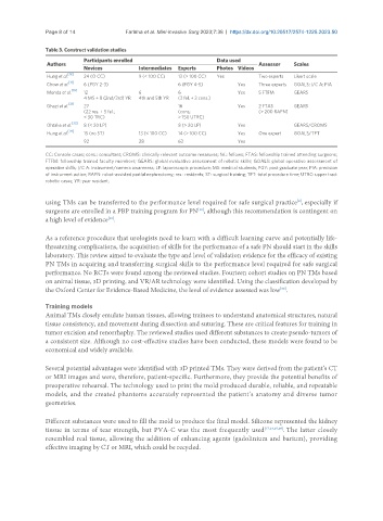
Table 3. Construct validation studies
Participants enrolled Data used
Authors Assessor Scales
Novices Intermediates Experts Photos Videos
Hung et al. [18] 24 (O CC) 9 (< 100 CC) 13 (> 100 CC) Yes Two experts Likert scale
[11]
Chow et al. 6 (PGY 2-3) 6 (PGY 4-5) Yes Three experts GOALS; I/C A; PIA
Monda et al. [19] 12 6 6 Yes 5 FTFM GEARS
4 MS + 8 (2nd/3rd) YR 4th and 5th YR (3 fel. + 3 cons.)
[21]
Ghazi et al. 27 16 Yes 2 FTAS GEARS
(22 res. + 5 fel.; (cons; (> 200 RAPN)
< 30 TRC) > 150 UTRC)
Ohtake et al. [33] 8 (< 20 LP) 8 (> 20 LP) Yes GEARS/CROMS
[29]
Hung et al. 15 (no ST) 13 (< 100 CC) 14 (> 100 CC) Yes One expert GOALS/TPT
92 28 63 Yes
CC: Console cases; cons.: consultant; CROMS: clinically relevant outcome measures; fel.: fellows; FTAS: fellowship trained attending surgeons;
FTFM: fellowship trained faculty members; GEARS: global evaluative assessment of robotic skills; GOALS: global operative assessment of
operative skills; I/C A: instrument/camera awareness; LP: laparoscopic procedure; MS: medical students; PGY: post graduate year; PIA: precision
of instrument action; RAPN: robot-assisted partial nephrectomy; res.: residents; ST: surgical training; TPT: total procedure time; UTRC: upper tract
robotic cases; YR: year resident.
using TMs can be transferred to the performance level required for safe surgical practice , especially if
[8]
[10]
surgeons are enrolled in a PBP training program for PN , although this recommendation is contingent on
a high level of evidence .
[10]
As a reference procedure that urologists need to learn with a difficult learning curve and potentially life-
threatening complications, the acquisition of skills for the performance of a safe PN should start in the skills
laboratory. This review aimed to evaluate the type and level of validation evidence for the efficacy of existing
PN TMs in acquiring and transferring surgical skills to the performance level required for safe surgical
performance. No RCTs were found among the reviewed studies. Fourteen cohort studies on PN TMs based
on animal tissue, 3D printing, and VR/AR technology were identified. Using the classification developed by
the Oxford Center for Evidence-Based Medicine, the level of evidence assessed was low .
[30]
Training models
Animal TMs closely emulate human tissues, allowing trainees to understand anatomical structures, natural
tissue consistency, and movement during dissection and suturing. These are critical features for training in
tumor excision and renorrhaphy. The reviewed studies used different substances to create pseudo-tumors of
a consistent size. Although no cost-effective studies have been conducted, these models were found to be
economical and widely available.
Several potential advantages were identified with 3D printed TMs. They were derived from the patient’s CT
or MRI images and were, therefore, patient-specific. Furthermore, they provide the potential benefits of
preoperative rehearsal. The technology used to print the mold produced durable, reliable, and repeatable
models, and the created phantoms accurately represented the patient’s anatomy and diverse tumor
geometries.
Different substances were used to fill the mold to produce the final model. Silicone represented the kidney
tissue in terms of tear strength, but PVA-C was the most frequently used [17,23,25,26] . The latter closely
resembled real tissue, allowing the addition of enhancing agents (gadolinium and barium), providing
effective imaging by CT or MRI, which could be recycled.