
Page 65 - Read Online
P. 65
Imai et al. Mini-invasive Surg 2024;8:2 https://dx.doi.org/10.20517/2574-1225.2023.67 Page 3 of 8
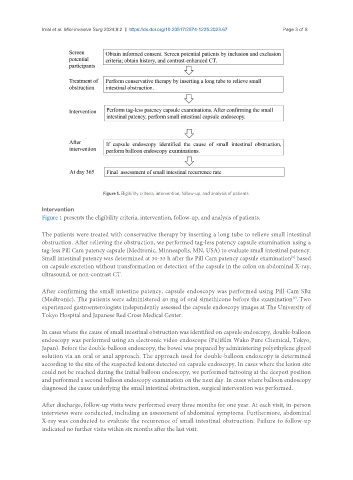
Figure 1. Eligibility criteria, intervention, follow-up, and analysis of patients.
Intervention
Figure 1 presents the eligibility criteria, intervention, follow-up, and analysis of patients.
The patients were treated with conservative therapy by inserting a long tube to relieve small intestinal
obstruction. After relieving the obstruction, we performed tag-less patency capsule examination using a
tag-less Pill Cam patency capsule (Medtronic, Minneapolis, MN, USA) to evaluate small intestinal patency.
Small intestinal patency was determined at 30-33 h after the Pill Cam patency capsule examination based
[8]
on capsule excretion without transformation or detection of the capsule in the colon on abdominal X-ray,
ultrasound, or non-contrast CT.
After confirming the small intestine patency, capsule endoscopy was performed using Pill Cam SB2
(Medtronic). The patients were administered 40 mg of oral simethicone before the examination . Two
[9]
experienced gastroenterologists independently assessed the capsule endoscopy images at The University of
Tokyo Hospital and Japanese Red Cross Medical Center.
In cases where the cause of small intestinal obstruction was identified on capsule endoscopy, double-balloon
endoscopy was performed using an electronic video endoscope (Fujifilm Wako Pure Chemical, Tokyo,
Japan). Before the double-balloon endoscopy, the bowel was prepared by administering polyethylene glycol
solution via an oral or anal approach. The approach used for double-balloon endoscopy is determined
according to the site of the suspected lesions detected on capsule endoscopy. In cases where the lesion site
could not be reached during the initial balloon endoscopy, we performed tattooing at the deepest position
and performed a second balloon endoscopy examination on the next day. In cases where balloon endoscopy
diagnosed the cause underlying the small intestinal obstruction, surgical intervention was performed.
After discharge, follow-up visits were performed every three months for one year. At each visit, in-person
interviews were conducted, including an assessment of abdominal symptoms. Furthermore, abdominal
X-ray was conducted to evaluate the recurrence of small intestinal obstruction. Failure to follow-up
indicated no further visits within six months after the last visit.