
Page 16 - Read Online
P. 16
Page 2 of 10 Lambert et al. J Transl Genet Genom 2018;2:11. I https://doi.org/10.20517/jtgg.2018.11
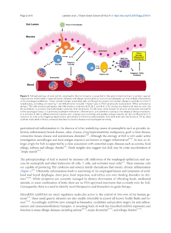
Figure 1. Pathophysiology of eosinophilic esophagitis. Barrier integrity is essential in the gastrointestinal tract to protect against
inappropriate inflammation. Impaired barrier defenses and allergic sensitization to food and aeroallergens can then initiate inflammation
in the esophageal epithelium. Tissue resident antigen-presenting cells are thought to process and present allergenic peptides to CD4+ T
lymphocytes, activating and skewing T cell differentiation towards T helper type 2 (Th2) lymphocyte development. When activated by
antigen, Th2 cells produce pathogenic and inflammatory cytokines, IL-13, IL-5, and IL-4. IL-13 induces non-traditional immune cells, like
the epithelium, to produce chemoattractant cytokines. One chemokine, CCL26 (also called Eotaxin-3) attracts and recruits eosinophils
to the esophageal tissue. IL-5 promotes eosinophil maturation, differentiation, and activation, leading to the release of allergic mediators.
IL-4 activates B cells to differentiate into plasma cells, undergo class switching, and produce antigen-specific IgE. IgE can then bind to Fc
receptors on mast cells, triggering degranulation and release of histamine and proteases. Activated mast cells also produce TGF-β, a key
cytokine implicated in fibrotic processes that lead to stenotic disease and esophageal narrowing
gastrointestinal inflammation in the absence of other underlying causes of eosinophilia such as parasitic in-
fection, inflammatory bowel disease, celiac disease, drug hypersensitivity, malignancy, graft-vs-host disease,
[7,8]
connective tissues disease and autoimmune disorders . Although the etiology of EoE is still under active
investigation, aeroallergen and food antigen exposure are known to trigger inflammation [9-11] . In fact, an al-
lergic origin for EoE is supported by a close association with comorbid atopic diseases such as eczema, food
[12]
allergy, asthma and allergic rhinitis . Fresh insights also suggest that EoE may be a late manifestation of
[13]
“atopic march” .
The pathophysiology of EoE is marked by immune cell infiltration of the esophageal epithelium and mu-
cosa by eosinophils and other leukocytes (B cells, T cells, and activated mast cells) . These immune cells
[14]
are capable of producing Th2 cytokines and eotaxin family chemokines that sustain chronic inflammation
[Figure 1] . Ultimately, inflammation leads to narrowing of the esophageal lumen and symptoms of solid-
[15]
food and liquid dysphagia, chest pain, food impaction, acid-reflux and even feeding disorders in chil-
dren [14,16] . While symptoms are currently managed by dietary elimination of offending foods, swallowed
steroids, or some combination of both, there are no FDA-approved treatments that currently exist for EoE.
Consequently, there is a need to identify novel therapeutics and biomarkers to guide therapy.
MicroRNA (miRNAs) are small regulatory molecules active in the control of 30%-60% of the human ge-
[17]
nome . These small genetic elements are also readily detectable in almost all known bodily fluids and tis-
sues [18,19] . Accordingly, miRNAs have emerged as biomarker candidates and putative targets for anti-inflam-
matory and immunomodulatory therapies. A mounting body of work has described miRNA expression and
function in many allergic diseases, including asthma [20-23] , atopic dermatitis [24,25] , and allergic rhinitis [21,26] .