
Page 1005 - Read Online
P. 1005
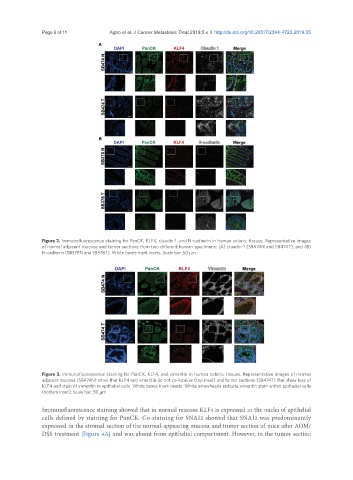
Page 6 of 11                                 Agbo et al. J Cancer Metastasis Treat 2019;5:x  I  http://dx.doi.org/10.20517/2394-4722.2019.35


               Figure 2. Immunofluorescence staining for PanCK, KLF4, claudin-1, and N-cadherin in human colonic tissues. Representative images
               of normal adjacent mucosa and tumor sections from two different human specimens: (A) claudin-1 (SB474N and SB474T); and (B)
               N-cadherin (SB378N and SB378T). White boxes mark insets. Scale bar: 50 μm


               Figure 3. Immunofluorescence staining for PanCK, KLF4, and vimentin in human colonic tissues. Representative images of normal
               adjacent mucosa (SB474N) show that KLF4 and vimentin do not co-localize (top inset) and tumor sections (SB474T) that show loss of
               KLF4 and stain of vimentin in epithelial cells. White boxes mark insets. White arrowheads indicate vimentin stain within epithelial cells
               (bottom inset). Scale bar: 50 μm

               Immunofluorescence staining showed that in normal mucosa KLF4 is expressed in the nuclei of epithelial
               cells defined by staining for PanCK. Co-staining for SNAI2 showed that SNAI2 was predominantly
               expressed in the stromal section of the normal-appearing mucosa and tumor section of mice after AOM/
               DSS treatment [Figure 4A] and was absent from epithelial compartment. However, in the tumor section