
Page 1004 - Read Online
P. 1004
Agbo et al. J Cancer Metastasis Treat 2019;5:x I http://dx.doi.org/10.20517/2394-4722.2019.35 Page 5 of 11
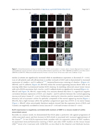
Figure 1. Immunofluorescence staining for PanCK, KLF4, TWIST, and β-catenin in human colonic tissues. Representative images of
normal adjacent mucosa and tumor sections from two different human specimens: (A) TWIST (SB396N and SB396T); and (B) β-catenin
(SB474N and SB474T). White arrowheads indicate KLF4 stain in stromal tissue. White boxes mark insets. Scale bar: 50 μm
nuclear β-catenin are significantly increased while its membranous expression is decreased (P < 0.001).
Loss of cell polarity and cell-cell junctions is another hallmark of EMT and is characterized by increased
expression of claudin-1 and N-cadherin [23-27] . Immunofluorescence staining of claudin-1 [Figure 2A,
SB474N] and N-cadherin [Figure 2B, SB378N] within the normal-appearing mucosa shows little or no
staining while there is pronounced nuclear KLF4 staining. In matching colorectal cancer tumor tissues
with lack of KLF4 expression, both claudin-1 and N-cadherin levels are significantly increased [Figure 2A,
SB474T and Figure 2B, SB378T, respectively]. Our analysis showed that there is a significant negative
correlation between expression of KLF4 and claudin-1 and N-cadherin between the normal-appearing
mucosa and tumor tissues (P < 0.001). Furthermore, staining for vimentin, another mesenchymal marker,
showed a lack of expression within the epithelial component of the normal-appearing mucosa [Figure 3,
SB474N], but a slight increase within the epithelial compartment upon loss of KLF4 in the tumor tissues
[Figure 3, SB474T, white arrowheads]. Statistical analysis showed that the expression levels of KLF4 and
vimentin are negatively correlated in normal mucosa (P < 0.05) and in tumor sections (P < 0.001).
KLF4 expression is negatively correlated with markers of EMT in a mouse model of colitis-
associated cancer
Recently, in a mouse model, we demonstrated that KLF4 plays a protective role against progression of
colitis-associated cancer and that decrease in KLF4 levels is associated with increased aggressiveness of
[18]
the disease . To test if KLF4 expression levels correlate with the expression pattern of EMT markers,
we performed immunofluorescence staining of KLF4 and select EMT markers on the mouse tissues
(Klf4 fl/fl mice) after treatment with AOM/DSS, as described in the Material and Methods Section.