
Page 276 - Read Online
P. 276
Finzel et al. J Cancer Metastasis Treat 2018;4:21 I http://dx.doi.org/10.20517/2394-4722.2018.10 Page 5 of 10
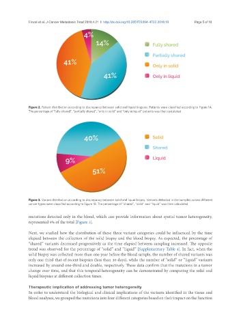
Figure 2. Patient distribution according to discrepancy between solid and liquid biopsies. Patients were classified according to Figure 1A.
The percentage of “fully shared”, “partially shared”, “only in solid” and “only in liquid” patients was then calculated
Figure 3. Variant distribution according to discrepancy between solid and liquid biopsy. Variants detected in the samples across different
cancer types were classified according to Figure 1B. The percentage of “shared”, “solid” and “liquid” was then calculated
mutations detected only in the blood, which can provide information about spatial tumor heterogeneity,
represented 9% of the total [Figure 3].
Next, we studied how the distribution of these three variant categories could be influenced by the time
elapsed between the collection of the solid biopsy and the blood biopsy. As expected, the percentage of
“shared” variants decreased progressively as the time elapsed between sampling increased. The opposite
trend was observed for the percentage of “solid” and “liquid” [Supplementary Table 4]. In fact, when the
solid biopsy was collected more than one year before the blood sample, the number of shared variants was
only one third that of recent biopsies (less than 30 days), while the number of “solid” or “liquid” variants
increased by around one-third and double, respectively. These data confirm that the mutations in a tumor
change over time, and that this temporal heterogeneity can be demonstrated by comparing the solid and
liquid biopsies at different collection times.
Therapeutic implication of addressing tumor heterogeneity
In order to understand the biological and clinical implications of the variants identified in the tissue and
blood analyses, we grouped the mutations into four different categories based on their impact on the function