
Page 278 - Read Online
P. 278
Finzel et al. J Cancer Metastasis Treat 2018;4:21 I http://dx.doi.org/10.20517/2394-4722.2018.10 Page 7 of 10
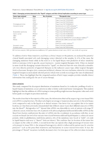
Table 1. Damaging variants detected in the “liquid” category and their clinical implication according to cancer type
Cancer type analysed Gene No. of variants Therapeutic value
Breast cancer HR+ ESR1 2 Resistance to aromatase inhibitors [10]
KRAS 1 Phase 2 trial (NCT02576444)
TP53 3 Phase 2 trial (NCT02576444)
Cholangiocarcinoma PIK3CA 1 Phase 2 trial (NCT02465060)
Colorectal cancer KRAS 4 Resistance to anti-EGFR antibodies [11]
PIK3CA 1 Resistance to anti-EGFR antibodies [12]
TP53 2 Phase 2 trial (NCT02576444)
Endometrial carcinoma TP53 1 Phase 2 trial (NCT02576444)
Gastric cancer PIK3CA 1 Phase 2 trial (NCT02465060)
GBM BRAF 1 Phase 2 trial (NCT02465060)
GIST KIT 1 Resistance to KIT/PDGFRA-tyrosine kinase inhibitors [13]
NSCLC EGFR 1 Resistance to EGFR-tyrosine kinase inhibitors [14]
GNAS 1 Unknown
MAP2K1 1 Sensitivity to MEK inhibitors [15]
Pancreatic cancer KRAS 1 Phase 2 trial (NCT02576444)
Prostate cancer TP53 2 Phase 2 trial (NCT02576444)
HR: hormone receptor; GBM: glioblastoma multiforme; GIST: gastrointestinal stromal tumor; NSCLC: non-small cell lung cancer
To address whether these mutations could have a clinical impact on the patients, we analysed the potential
clinical benefit associated with each damaging variant detected in the samples. In 97% of the cases, the
damaging mutations found solely in the solid or in the liquid biopsy were predictive of either sensitivity
(82%) or resistance (15%) to specific cancer treatments - mainly targeted therapies (98%). When we studied
in more detail the damaging variants detected in “liquid”, we observed that 96% were clinically actionable:
42% were directly predictive of approved therapies in the indicated cancer type, either targeted therapies
(80%) or hormone therapies (20%, aromatase inhibitors), whereas 54% were inclusion criteria for trials using
targeted therapies in molecularly selected patients (which were actively recruiting at the time of submission)
[Table 1]. These data highlight that the integrated analysis of both biopsy samples provides valuable clinical
information that could guide the use of cancer therapy.
DISCUSSION
This study compared the discrepant distribution of mutations found in the molecular profiling of solid vs.
liquid biopsies of metastatic cancer patients in order to better understand tumor heterogeneity. This analysis
highlighted that the addition of ctDNA testing to tissue profiling might increase therapeutic value and could
better guide oncologists in precision medicine.
The results show that in the majority of the cases, the information obtained by sequencing tumor tissue DNA
and ctDNA is complimentary. We observed a higher percentage of mutations detected only in the solid biopsy
(51%) compared to only in the liquid or to shared variants. One factor that can explain this is the tumor
location, as it has been previously demonstrated that different cancer types shed different amounts of DNA
into the blood . Bettegowda et al. showed that ctDNA was detectable in 100% of patients with stage IV
[16]
[16]
colorectal cancer, while less than 10% of patients with advanced gliomas harboured detectable ctDNA (as the
blood-brain barrier could prevent the entry of ctDNA into the circulation). In accordance with this, in our
analysis we found that 56% of the variants were shared between solid and liquid biopsies in colorectal cancer
patients, while in glioblastoma multiforme patients, 89% of the mutations were found in “solid”, 11% only
in liquid biopsies and none in “shared” (data not shown). This confirms that the location of the tumor has
an impact on the utility of ctDNA. A second factor that can explain why a high percentage of variants were
only present in tissue DNA is the temporal heterogeneity, which is influenced by patient-specific selective
pressures such as the prescribed treatments and fluctuations in tumor microenvironment. In fact, the
[17]
percentage of “shared” mutations markedly decreased when the time space of the collection dates increased.