Page 282 - Read Online
P. 282
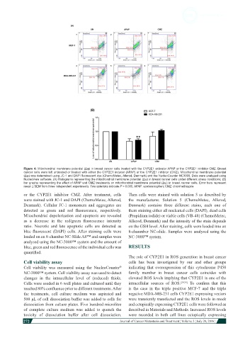
Figure 4: Mitochondrial membrane potential (Δѱ) in breast cancer cells treated with the CYP2E1 activator APAP or the CYP2E1 inhibitor CMZ. Breast
            cancer cells were left untreated or treated with either the CYP2E1 inducer (APAP) or the CYP2E1 inhibitor (CMZ). Mitochondrial membrane potential
            (Δѱ) was determined using JC-1 and DAPI fluorescent dye (ChemoMetec, Allerod, Denmark) and the NucleoCounter NC3000. Data were analyzed using
            NucleoView software. (A) Histograms representing the mitochondrial membrane potential (Δѱ) in breast cancer cells under different stress conditions; (B)
            bar graphs representing the effect of APAP and CMZ treatments on mitochondrial membrane potential (Δѱ) in breast cancer cells. Error bars represent
            mean ± SEM from three independent experiments. Two asterisks indicate P < 0.005. APAP: acetaminophen; CMZ: chlormethiazole
            or  the  CYP2E1  inhibitor  CMZ.  After  treatment,  cells   Then cells were stained with solution 5 as described by
            were stained with JC-1 and DAPI (ChemoMetec, Allerod,   the  manufacturer. Solution  5 (ChemoMetec,  Allerod,
            Denmark).  Cellular  JC-1 monomers  and aggregates  are   Denmark)  contains three  different  stains,  each  one  of
            detected  as  green  and  red  fluorescence,  respectively.   them staining either all nucleated cells (DAPI), dead cells
            Mitochondrial depolarization and apoptosis are revealed   (Propidium iodide) or viable cells (VB-48) (ChemoMetec,
            as  a  decrease  in  the  red/green  fluorescence  intensity   Allerod, Denmark) and the intensity of the stain depends
            ratio.  Necrotic  and  late  apoptotic  cells  are  detected  as   on the GSH level. After staining, cells were loaded into an
            blue  fluorescent  (DAPI)  cells. After  staining  cells  were   8-chanmber NC-slide. Samples were analysed using the
            loaded on an 8-chamber NC-Slide A8™ and samples were   NC-3000™ system.
            analysed using the NC-3000™ system and the amount of
            blue, green and red fluorescence of the individual cells was   RESULTS
            quantified.
                                                              The role of CYP2E1 in ROS generation in breast cancer
            Cell viability assay                              cells  has been  investigated  by our and  other  groups
            Cell viability  was measured using the NucleoCounter    indicating  that  overexpression  of this  cytochrome  P450
                                                          ®
            NC-3000™ system. Cell viability assay was used to detect   family  member in  breast  cancer  cells  coincides  with
            changes  in  the  intracellular  level  of (reduced)  thiols.   elevated ROS levels implying that CYP2E1 is one of the
            Cells were seeded in 6 well plates and cultured until they   intracellular  sources of ROS. [29,34]   To  confirm  that  this
            reached 80% confluence prior to different treatments. After   is the  case  in the  triple  positive  MCF-7 and  the  triple
            the treatments, cell culture medium was aspirated and   negative MDA-MB-231 cells CYP2E1 expressing vectors
            500 μL of cell dissociation buffer was added to cells for   were transiently transfected and the ROS levels in mock
            dissociation from culture plates. Five hundred microlitre   and ectopically expressing CYP2E1 cells were followed as
            of complete  culture  medium  was added  to quench  the   described in Materials and Methods. Increased ROS levels
            toxicity  of dissociation  buffer after  cell  dissociation.   were  recorded  in both  cell  lines  ectopically  expressing
            272
                                                                                                                        Journal of Cancer Metastasis and Treatment ¦ Volume 2 ¦ July 29, 2016 ¦

