
Page 274 - Read Online
P. 274
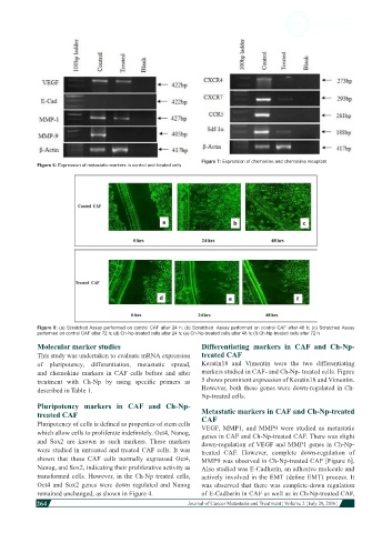
Figure 7: Expression of chemokine and chemokine receptors
            Figure 6: Expression of metastatic markers in control and treated cells


            Figure 8: (a) Scratched Assay performed on control CAF after 24 h; (b) Scratched  Assay performed on control CAF after 48 h; (c) Scratched Assay
            performed on control CAF after 72 h; (d) Ch-Np-treated cells after 24 h; (e) Ch-Np-treated cells after 48 h; (f) Ch-Np-treated cells after 72 h
            Molecular marker studies                          Differentiating markers in CAF and  Ch-Np-
            This study was undertaken to evaluate mRNA expression   treated CAF
            of pluripotency, differentiation, metastatic spread,   Keratin18  and  Vimentin  were  the  two  differentiating
            and  chemokine  markers  in  CAF  cells  before  and  after   markers studied in CAF- and Ch-Np- treated cells. Figure
            treatment  with  Ch-Np  by  using  specific  primers  as   5 shows prominent expression of Keratin18 and Vimentin.
            described in Table 1.                             However, both these genes were down-regulated in Ch-
                                                              Np-treated cells.
            Pluripotency markers in CAF and Ch-Np-
            treated CAF                                       Metastatic markers in CAF and Ch-Np-treated
                                                              CAF
            Pluripotency of cells is defined as properties of stem cells   VEGF,  MMP1,  and  MMP9  were  studied  as  metastatic
            which allow cells to proliferate indefinitely. Oct4, Nanog,   genes in CAF and Ch-Np-treated CAF. There was slight
            and Sox2 are known as such markers.  These markers   down-regulation of VEGF and MMP1 genes in Ch-Np-
            were studied in untreated and treated CAF cells. It was   treated  CAF.  However,  complete  down-regulation  of
            shown  that  these  CAF  cells  normally  expressed  Oct4,   MMP9 was observed in Ch-Np-treated CAF [Figure 6].
            Nanog, and Sox2, indicating their proliferative activity as   Also studied was E-Cadherin, an adhesive molecule and
            transformed cells. However, in the Ch-Np treated cells,   actively involved in the EMT (define EMT) process. It
            Oct4  and  Sox2  genes  were  down  regulated  and  Nanog   was observed that there was complete-down regulation
            remained unchanged, as shown in Figure 4.         of E-Cadherin in CAF as well as in Ch-Np-treated CAF,
            264
                                                                                                                        Journal of Cancer Metastasis and Treatment ¦ Volume 2 ¦ July 29, 2016 ¦