
Page 272 - Read Online
P. 272
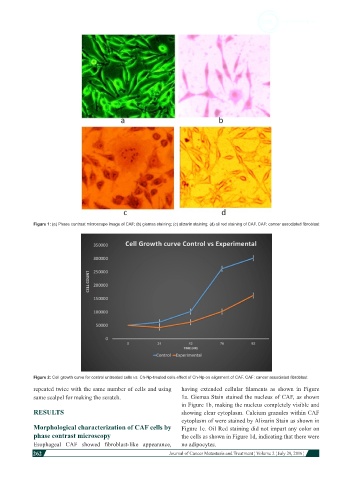
Figure 1: (a) Phase contrast microscope image of CAF; (b) giemsa staining; (c) alizarin staining; (d) oil red staining of CAF. CAF: cancer associated fibroblast


            Figure 2: Cell growth curve for control untreated cells vs. Ch-Np-treated cells effect of Ch-Np on alignment of CAF. CAF: cancer associated fibroblast
            repeated twice with the same number of cells and using   having  extended  cellular  filaments  as  shown  in  Figure
            same scalpel for making the scratch.              1a. Giemsa Stain stained the nucleus of CAF, as shown
                                                              in Figure 1b, making the nucleus completely visible and
            RESULTS                                           showing clear cytoplasm. Calcium granules within CAF
                                                              cytoplasm of were stained by Alizarin Stain as shown in
            Morphological characterization of CAF cells by    Figure 1c. Oil Red staining did not impart any color on
            phase contrast microscopy                         the cells as shown in Figure 1d, indicating that there were
            Esophageal  CAF  showed  fibroblast-like  appearance,   no adipocytes.
            262
                                                                                                                        Journal of Cancer Metastasis and Treatment ¦ Volume 2 ¦ July 29, 2016 ¦