
Page 25 - Read Online
P. 25
Page 4 of 16 Merhi et al. J Cancer Metastasis Treat 2021;7:42 https://dx.doi.org/10.20517/2394-4722.2021.80
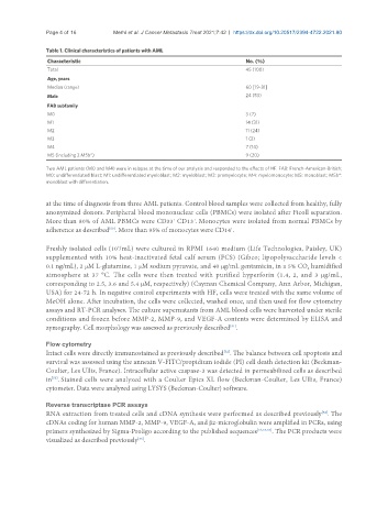
Table 1. Clinical characteristics of patients with AML
Characteristic No. (%)
Total 45 (100)
Age, years
Median (range) 60 [19-81]
Male 24 (53)
FAB subfamily
M0 3 (7)
M1 14 (31)
M2 11 (24)
M3 1 (2)
M4 7 (14)
M5 (including 2 M5b*) 9 (20)
Two AML patients (M0 and M4) were in relapse at the time of our analysis and responded to the effects of HF. FAB: French-American-British;
M0: undifferentiated blast; M1: undifferentiated myeloblast; M2: myeloblast; M3: promyelocyte; M4: myelomonocyte; M5: monoblast; M5b*:
monoblast with differentiation.
at the time of diagnosis from three AML patients. Control blood samples were collected from healthy, fully
anonymized donors. Peripheral blood mononuclear cells (PBMCs) were isolated after Ficoll separation.
+
+
More than 80% of AML PBMCs were CD33 CD13 . Monocytes were isolated from normal PBMCs by
adherence as described . More than 95% of monocytes were CD14 .
+
[50]
Freshly isolated cells (10 /mL) were cultured in RPMI 1640 medium (Life Technologies, Paisley, UK)
6
supplemented with 10% heat-inactivated fetal calf serum (FCS) (Gibco; lipopolysaccharide levels <
0.1 ng/mL), 2 μM L-glutamine, 1 μM sodium pyruvate, and 40 μg/mL gentamicin, in a 5% CO humidified
2
atmosphere at 37 °C. The cells were then treated with purified hyperforin (1.4, 2, and 3 μg/mL,
corresponding to 2.5, 3.6 and 5.4 μM, respectively) (Cayman Chemical Company, Ann Arbor, Michigan,
USA) for 24-72 h. In negative control experiments with HF, cells were treated with the same volume of
MeOH alone. After incubation, the cells were collected, washed once, and then used for flow cytometry
assays and RT-PCR analyses. The culture supernatants from AML blood cells were harvested under sterile
conditions and frozen before MMP-2, MMP-9, and VEGF-A contents were determined by ELISA and
zymography. Cell morphology was assessed as previously described .
[51]
Flow cytometry
Intact cells were directly immunostained as previously described . The balance between cell apoptosis and
[52]
survival was assessed using the annexin V-FITC/propidium iodide (PI) cell death detection kit (Beckman-
Coulter, Les Ullis, France). Intracellular active caspase-3 was detected in permeabilized cells as described
in . Stained cells were analyzed with a Coulter Epics XL flow (Beckman-Coulter, Les Ullis, France)
[53]
cytometer. Data were analyzed using LYSYS (Beckman-Coulter) software.
Reverse transcriptase PCR assays
RNA extraction from treated cells and cDNA synthesis were performed as described previously . The
[52]
cDNAs coding for human MMP-2, MMP-9, VEGF-A, and β2-microglobulin were amplified in PCRs, using
primers synthesized by Sigma-Proligo according to the published sequences [52,54,55] . The PCR products were
[56]
visualized as described previously .